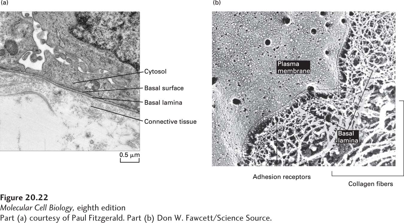
FIGURE 20- 22 A basal lamina separates epithelial cells and some other cells from connective tissue. (a) Transmission electron micrograph of a thin section of cells (top) and underlying connective tissue (bottom). The electron- dense layer of the basal lamina can be seen to follow the undulations of the basal surfaces of the cells. (b) Electron micrograph of a quick- freeze deep- etch preparation of skeletal muscle, showing the plasma membrane, basal lamina, and surrounding connective- tissue collagen fibers. In this preparation, the basal lamina is revealed as a meshwork of filamentous proteins that associates with the plasma membrane and the thicker collagen fibers of the connective tissue.
[Part (a) courtesy of Paul Fitzgerald. Part (b) Don W. Fawcett/Science Source.]
[Leave] [Close]