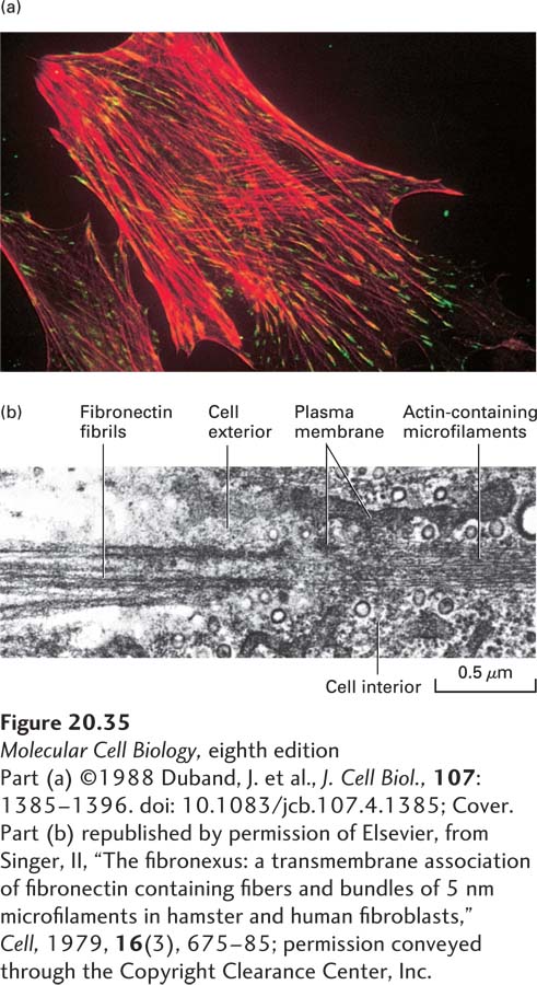
EXPERIMENTAL FIGURE 20- 35 Integrins mediate linkage between fibronectin in the ECM and the cytoskeleton. (a) Immunofluorescent micrograph of a fixed cultured fibroblast showing colocalization of the α5β1 integrin (green) and actin- containing stress fibers (red). The cell was incubated with two types of monoclonal antibodies: an integrin- specific antibody linked to a green- fluorescing dye and an actin- specific antibody linked to a red- fluorescing dye. Stress fibers are long bundles of actin microfilaments that radiate inward from points where the cell contacts a substratum. At the distal ends of these fibers, near the plasma membrane, the coincidence of actin (red) and fibronectin- binding integrin (green) produces a yellow fluorescence. (b) Electron micrograph of the junction of fibronectin and actin fibers in a cultured fibroblast. Individual actin- containing 7- nm microfilaments, components of a stress fiber, end at the obliquely sectioned cell membrane. The microfilaments appear aligned with and in close proximity to the thicker, densely stained fibronectin fibrils on the outside of the cell.
[Part (a) ©1988 Duband, J. et al., J. Cell Biol., 107:1385– 1396. doi: 10.1083/jcb.107.4.1385; Cover. Part (b) republished by permission of Elsevier, from Singer, II, “The fibronexus: a transmembrane association of fibronectin- containing fibers and bundles of 5 nm microfilaments in hamster and human fibroblasts,” Cell, 1979, 16(3), 675– 85; permission conveyed through the Copyright Clearance Center, Inc.]
[Leave] [Close]