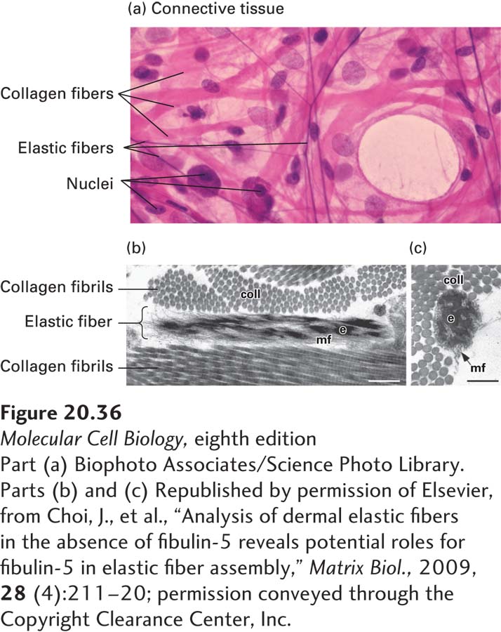
FIGURE 20- 36 Elastic and collagen fibers in connective tissue. (a) Light- microscopic image of loose connective tissue from the lung. Elastic fibers are the thin fibers that are stained purple, collagen fibers (bundles of collagen fibrils) are stained pink, and the nuclei of cells are stained purple. (b) Longitudinal and (c) cross- sectional electron microscopic images of elastic fibers and collagen fibrils (coll) in the skin of a mouse. The elastic fibers have a solid core of elastin (e) integrated into and surrounded by a bundle of microfibrils (mf). Scale bars, 0.25 μm.
[Part (a) Biophoto Associates/Science Photo Library. Parts (b) and (c) Republished by permission of Elsevier, from Choi, J., et al., “Analysis of dermal elastic fibers in the absence of fibulin- 5 reveals potential roles for fibulin- 5 in elastic fiber assembly,” Matrix Biol., 2009, 28 (4):211– 20; permission conveyed through the Copyright Clearance Center, Inc.]
[Leave] [Close]