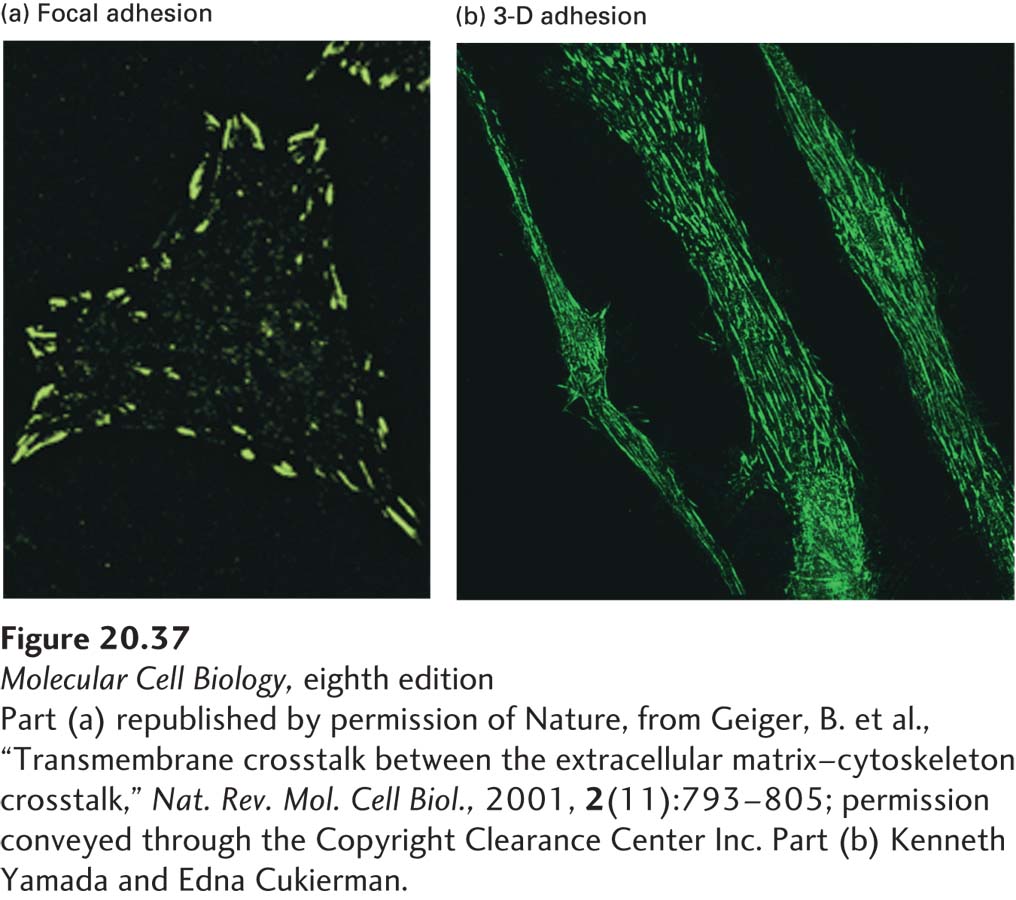
EXPERIMENTAL FIGURE 20- 37 Integrins cluster into adhesive structures with various morphologies in nonepithelial cells. Immunofluorescence methods were used to detect integrin- containing adhesive structures (green) on cultured cells. Shown here are (a) focal adhesions and (b) 3- D adhesions on the surfaces of human fibroblasts. Cells were grown (a) directly on the flat surface of a culture dish or (b) on a three- dimensional matrix of ECM components. The shape, distribution, and composition of the integrin- based adhesions formed by cells vary depending on the cells’ environment.
[Part (a) republished by permission of Nature, from Geiger, B. et al., “Transmembrane crosstalk between the extracellular matrix– cytoskeleton crosstalk,” Nat. Rev. Mol. Cell Biol., 2001, 2(11):793– 805; permission conveyed through the Copyright Clearance Center Inc. Part (b) Kenneth Yamada and Edna Cukierman.]
[Leave] [Close]