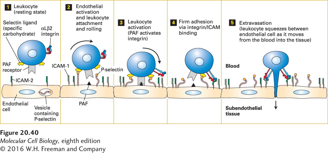
FIGURE 20- 40 Endothelium- leukocyte interactions: activation, binding, rolling, and extravasation. Step 1: In the absence of inflammation or infection, leukocytes and endothelial cells lining blood vessels are in a resting state and not interacting. Step 2: Inflammatory signals released only in areas of inflammation, infection, or both activate resting endothelial cells, resulting in the movement of vesicle- sequestered selectins to the cell surface. The exposed selectins mediate weak binding of leukocytes by interacting with carbohydrate ligands on leukocytes. Blood flow forces the loosely bound leukocytes to roll along the endothelial surface of the blood vessel (curved arrow). Activation of the endothelium also causes synthesis of platelet- activating factor (PAF) and ICAM- 1, both expressed on the endothelial cell surface. PAF and other, usually secreted, activators, including chemokines, then induce changes in the shapes of the leukocytes and activation of leukocyte integrins such as αLβ2, which is expressed by T lymphocytes (step 3). The subsequent tight binding between activated integrins on leukocytes and CAMs on the endothelium (e.g., ICAM- 2 and ICAM- 1) results in firm adhesion (step 4) and subsequent movement (extravasation) into the underlying tissue (step 5). See R. O. Hynes and A. Lander, 1992, Cell 68:303.
[Leave] [Close]