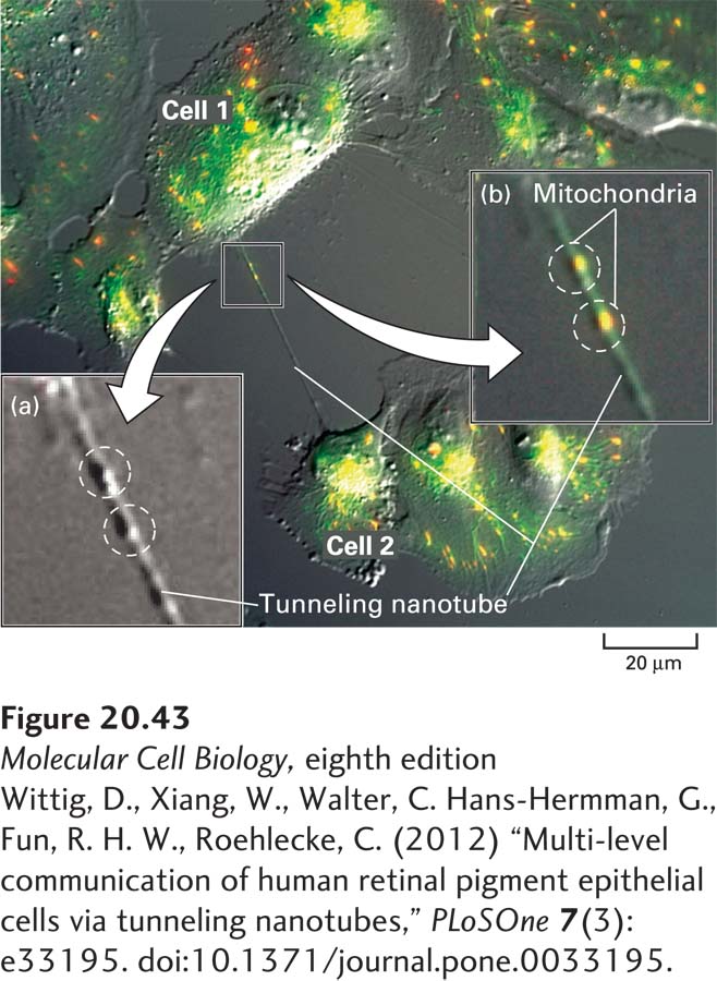
EXPERIMENTAL FIGURE 20- 43 Microscopic visualization of a tunneling nanotube and mitochondria in cultured human cells. Cultured human retinal pigment epithelial cells (ARPE- 19 cell line) were incubated with a fluorescent dye (JC- 1) that specifically stains mitochondria and then examined by a combination of conventional bright- field microscopy (see Chapter 4) to visualize the cells and fluorescence microscopy to visualize mitochondria (green intracellular fluorescence). A typical tunneling nanotube can be seen connecting cells 1 and 2. Inset (a) shows a higher magnification of the bright- field- only image with two bulges in the tunneling nanotube highlighted by dashed circles. Inset (b) shows a higher magnification of the same region of the combination image indicating two likely mitochondria within the tunneling nanotube at the positions of those bulges.
[Wittig, D., Xiang, W., Walter, C. Hans- Hermman, G., Fun, R. H. W., Roehlecke, C. (2012) “Multi- level communication of human retinal pigment epithelial cells via tunneling nanotubes,” PLoSOne 7(3): e33195. doi:10.1371/journal.pone.0033195.]
[Leave] [Close]