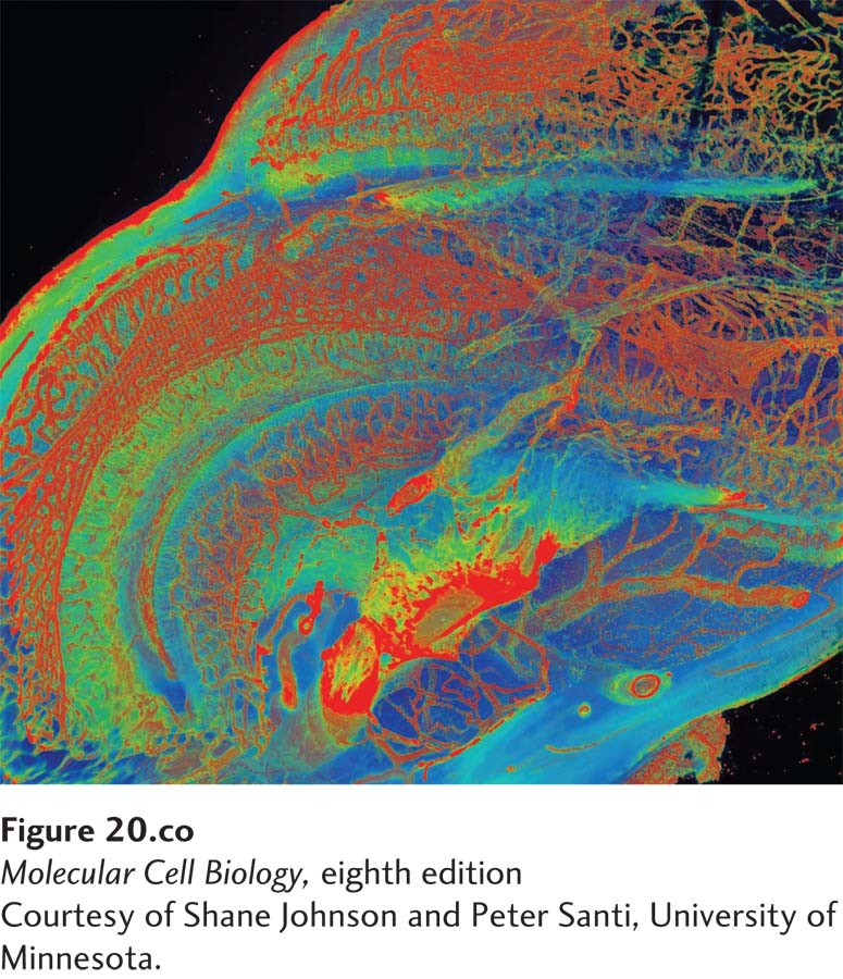
The cochlea of the inner ear uses mechanotransduction to convert the energy in sound waves into neuronal signals. The distribution of type IV collagen in the extracellular matrix of the cochlear duct of a mouse was visualized by scanning thin- sheet laser imaging microscopy after removing the cells with the detergent SDS and the calcium with the chelator EDTA. The sample was then stained first with an anti– type IV collagen antibody and then a fluorescently labeled secondary antibody. The false colors in the image represent the relative intensities of fluorescence (red > yellow > blue), and thus the relative local amounts of type IV collagen, in the basal lamina of the blood vessels (red), other basement membranes (yellow), and the cochlear wall (blue).
[Courtesy of Shane Johnson and Peter Santi, University of Minnesota.]
[Leave] [Close]