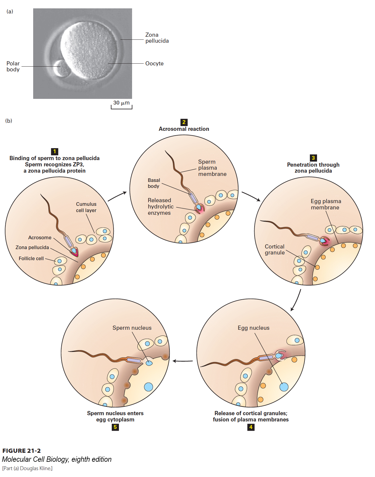
FIGURE 21- 2 Gamete fusion during fertilization. (a) Mammalian eggs, such as the mouse oocyte shown here, are surrounded by a ring of translucent material, the zona pellucida, which provides a binding matrix for sperm. The diameter of a mouse egg is ~70 µm, and the zona pellucida is ~6 µm thick. The polar body is a nonfunctional product of meiosis. (b) In the initial stage of fertilization (step 1), the sperm penetrates a layer of cumulus cells surrounding the egg to reach the zona pellucida. Interactions between GalT, a protein on the sperm surface, and ZP3, a glycoprotein in the zona pellucida, trigger the acrosomal reaction (step 2), which releases enzymes from the acrosome. Degradation of the zona pellucida by hydrolases and proteases released by the acrosomal reaction allows the sperm to begin entering the egg (step 3). Specific recognition proteins on the surfaces of egg and sperm facilitate fusion of their plasma membranes. Membrane fusion and the subsequent entry of the first sperm nucleus into the egg cytoplasm (steps 4 and 5) trigger the release of Ca2+ within the oocyte. Cortical granules (orange) respond to the Ca2+ surge by fusing with the oocyte membrane and releasing enzymes that act on the zona pellucida to prevent binding of additional sperm.
[Part (a) Douglas Kline.]
[Leave] [Close]