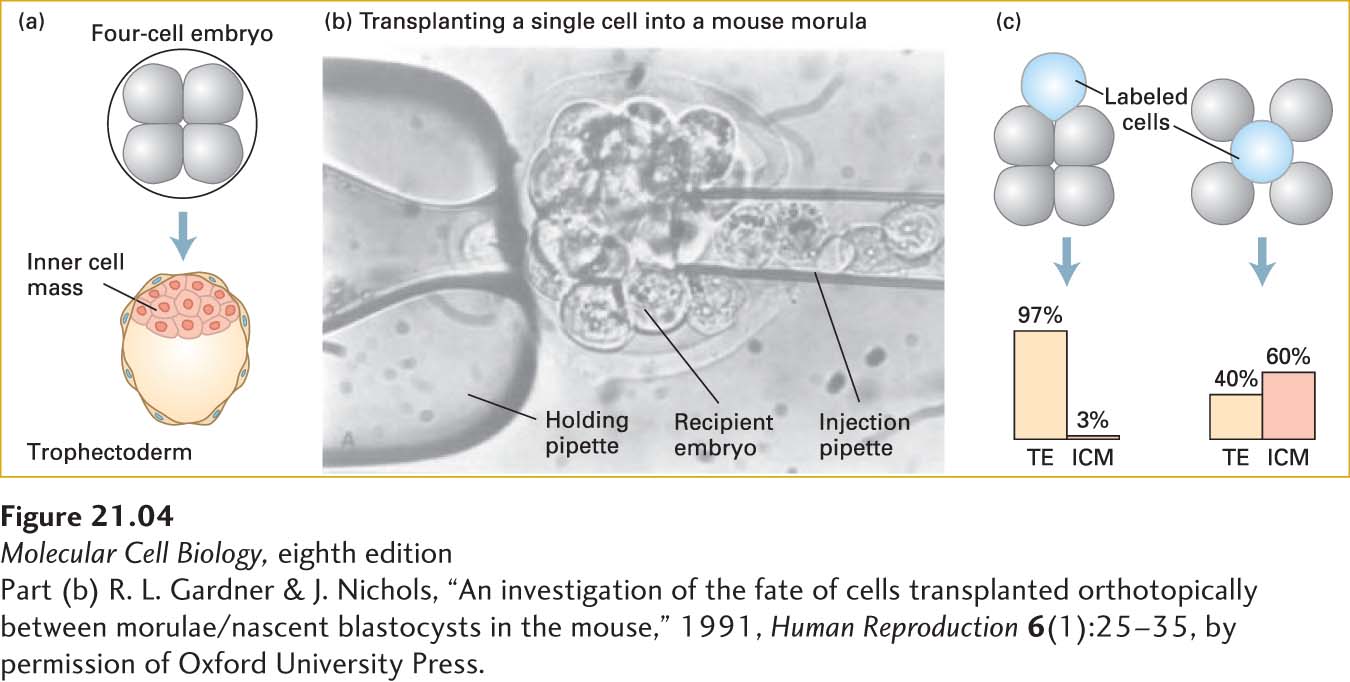
EXPERIMENTAL FIGURE 21- 4 Cell location determines cell fate in the early embryo. (a) A four- cell embryo normally develops into a blastocyst consisting of trophectoderm (TE) cells on the outside and inner cell mass (ICM) cells inside. (b) In order to discover whether position affects the fates of cells, transplantation experiments were done with mouse embryos. First, recipient morula- stage embryos had cells removed to make room for implanted cells. Then donor morula- stage (sixteen- cell) embryos were soaked in a dye that does not transfer between cells. Finally, labeled cells from the donor embryos were injected into inner or outer regions of the recipient embryos, as shown in the micrograph. The recipient embryo was held in place by a slight vacuum applied to the holding pipette. (c) The subsequent fates of the descendants of the transplanted labeled cells were monitored. For simplicity, four- cell recipient embryos are depicted, although morula- stage embryos were used as both donors and recipients. The results, summarized in the graphs, show that outer cells overwhelmingly form trophectoderm and that inner cells tend to become part of the ICM, but also form considerable trophectoderm.
[Part (b) R. L. Gardner & J. Nichols, “An investigation of the fate of cells transplanted orthotopically between morulae/nascent blastocysts in the mouse,” 1991, Human Reproduction 6(1):25– 35, by permission of Oxford University Press.]
[Leave] [Close]