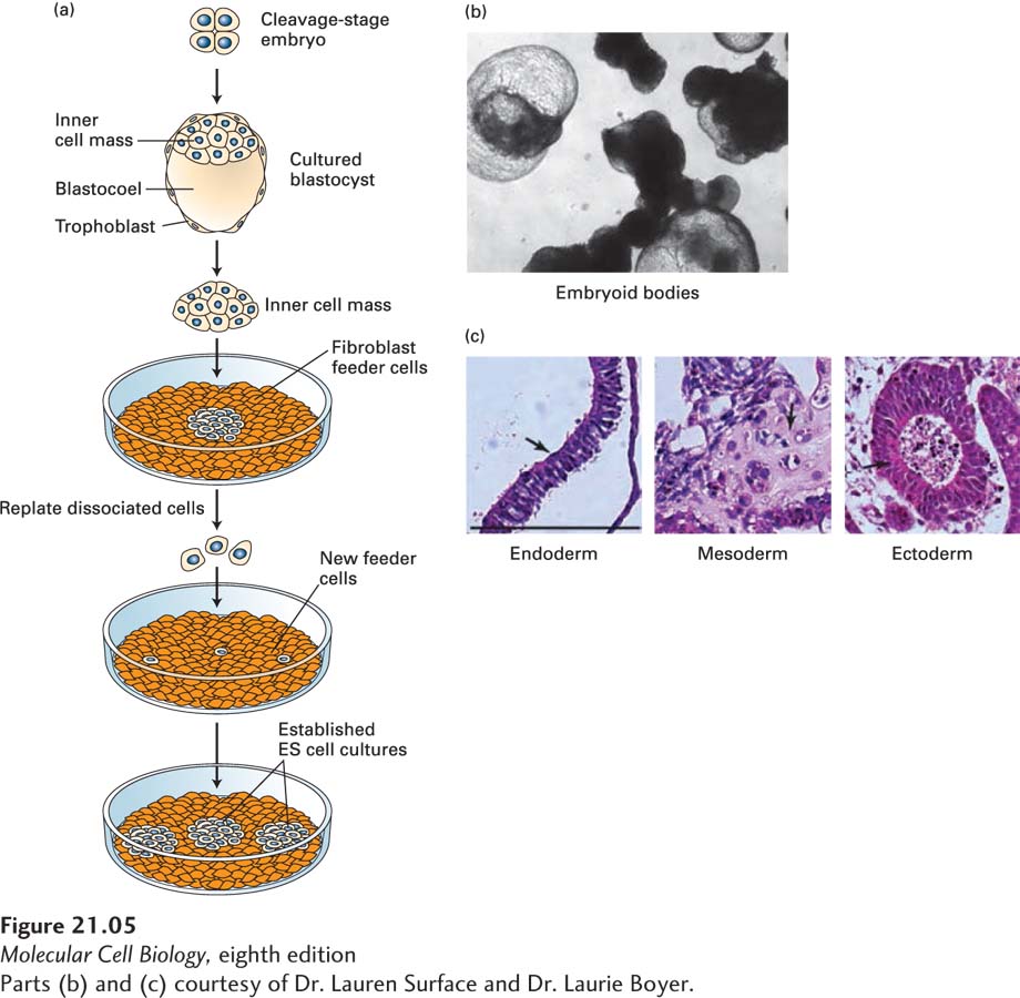
EXPERIMENTAL FIGURE 21- 5 Embryonic stem (ES) cells can be maintained in culture and can form differentiated cell types. (a) Human or mouse blastocysts are grown from cleavage- stage embryos produced by in vitro fertilization. The ICM is separated from the surrounding extraembryonic tissues and plated onto a layer of fibroblast cells, which help to nourish the embryonic cells by providing specific protein hormones. When individual cells are replated, they form colonies of ES cells, which can be maintained for many generations and can be stored frozen. ES cells can also be cultured without a fibroblast feeder layer if specific cytokines are added; leukemia inhibitory factor (LIF), for instance, supports growth of mouse ES cells by triggering activation of the Stat3 transcription factor; see J. S. Odorico et al., 2001, Stem Cells 19:193. (b) Embryonic stem cells allowed to differentiate in suspension culture become multicellular aggregates termed embryoid bodies. (c) Hematoxylin- and eosin- stained sections of embryoid bodies that contain derivatives of all three germ layers that are formed from the ICM during embryogenesis. Arrows in the images point to the following tissue types: (left) gut epithelium (endoderm), (middle) cartilage (mesoderm), and (right) neuroepithelial rosettes (ectoderm). Black bar = 100 µm.
[Parts (b) and (c) courtesy of Dr. Lauren Surface and Dr. Laurie Boyer.]
[Leave] [Close]