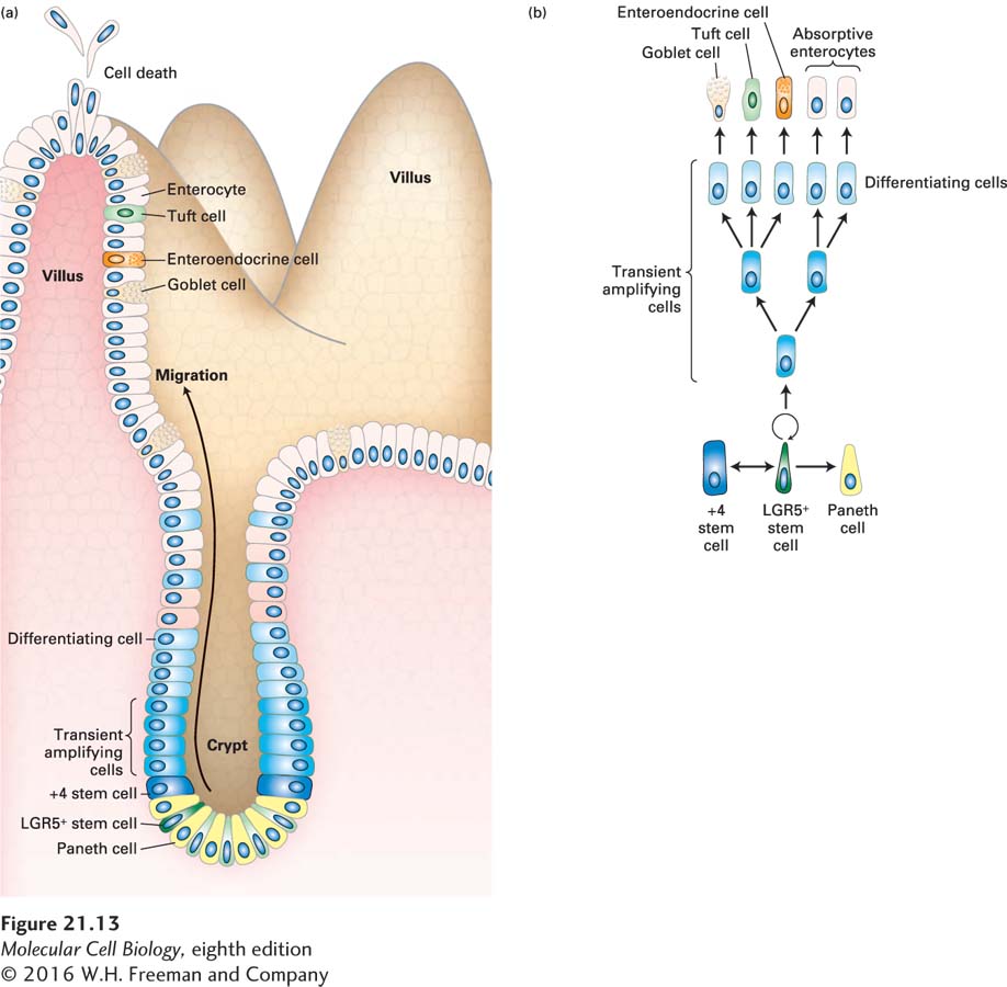
FIGURE 21- 13 Intestinal stem cells and their niche. (a) Schematic drawing of an intestinal crypt and villus, showing the Lgr5- expressing (Lgr5+) intestinal stem cells (dark green), their mitotic progeny, the transient amplifying cells (intermediate blue), the terminal differentiating cells (light blue), and the several types of differentiated cells in the villus. The base of the crypt is the location of Paneth cells (yellow), which provide a major part of the stem- cell niche and also secrete antimicrobial defense proteins. The +4 “reserve” stem cells (which occupy the fourth position from the crypt base, dark blue) can restore the Lgr5+ stem- cell compartment following injury and can also be generated from these stem cells. (b) Lineages of cells in the small intestine. Epithelial turnover occurs every 3– 5 days. New Paneth cells are supplied from the transient amplifying cells every 3– 6 weeks. See N. Barker, 2014, Nat. Rev. Mol. Cell Biol. 15:19.
[Leave] [Close]