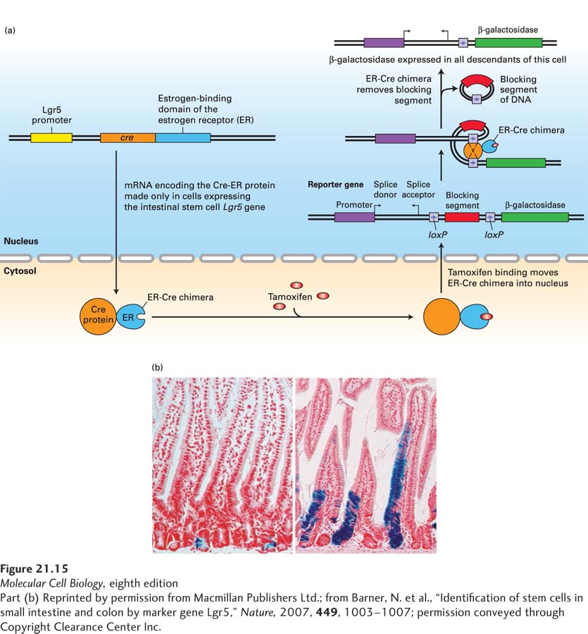
EXPERIMENTAL FIGURE 21- 15 Lineage- tracing studies show that the Lgr5- expressing cells at the bases of crypts are the intestinal stem cells. (a) Outline of the experiment. Using genetically altered ES cells (see Figure 6- 37 ), investigators generated one strain of mice in which a version of the gene encoding Cre recombinase (see Figure 6- 39 ) was placed under the control of the Lgr5 promoter, and thus Cre recombinase was produced only in cells, such as intestinal stem cells, that express the Lgr5 gene. This version of Cre recombinase contained an additional domain from the estrogen receptor (ER) that binds the estrogen analog tamoxifen; like the estrogen receptor and other nuclear receptors (see Figure 9- 45 ), the ER- Cre chimera is retained in the cytosol unless tamoxifen is added. In the presence of tamoxifen, ER- Cre moves into the nucleus, where it can interact with loxP sites in the chromosomal DNA. A second reporter strain of mice contained a bacterial β-galactosidase reporter gene that was preceded by two loxP sites. The blocking segment of DNA in between these loxP sites prevented expression of the β-galactosidase gene, and the β-galactosidase gene could be expressed only in cells where an active Cre recombinase had removed the sequence in between the two loxP sites. The two strains of mice were mated, and offspring containing both marker transgenes were identified. In these mice, β-galactosidase was expressed only in cells in which the Lgr5- controlled ER- Cre gene was expressed, and only after the estrogen analog tamoxifen was given to the mice. Thus only Lgr5- expressing cells— and all of their descendants— would express the β-galactosidase gene. (b) Results of the experiment. One day after tamoxifen was given to these mice, the only cells expressing β-galactosidase (indicated by the blue histochemical stain) were the Lgr5- expressing intestinal stem cells at the bases of the crypts (left). Five days after tamoxifen administration, additional blue cells— the epithelial descendants of the intestinal stem cells— were seen migrating up the sides of the villi. Some blue stem cells remained at the bottom of the crypt.
[Part (b) Reprinted by permission from Macmillan Publishers Ltd.; from Barner, N. et al., “Identification of stem cells in small intestine and colon by marker gene Lgr5,” Nature, 2007, 449, 1003– 1007; permission conveyed through Copyright Clearance Center Inc.]
[Leave] [Close]