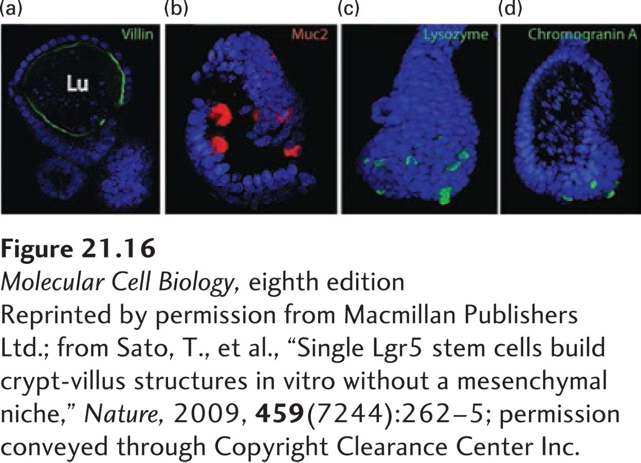
EXPERIMENTAL FIGURE 21- 16 Single Lgr5- expressing intestinal stem cells build crypt- villus structures in culture without niche cells. Single Lgr5- expressing cells isolated from intestinal crypts were placed in culture on a type IV extracellular matrix (see Figure 20- 23 ), the type of matrix that normally underlies and supports sheets of epithelial cells. After 2 weeks, these cultures had formed epithelial sheets that resembled villi in structure. Staining of these organoids for specific marker proteins showed that they contained all four differentiated epithelial cell types: (a) villin (green) is a marker protein for the absorptive enterocytes that are localized near the apical (luminal, Lu) surface of these organoids; (b) Muc2 (red) for goblet cells; (c) lysozyme (green) for Paneth cells; and (d) chromogranin A (green) for enteroendocrine cells. The organoids were also stained with DAPI (blue) to reveal nuclei.
[Reprinted by permission from Macmillan Publishers Ltd.; from Sato, T., et al., “Single Lgr5 stem cells build crypt- villus structures in vitro without a mesenchymal niche,” Nature, 2009, 459(7244):262– 5; permission conveyed through Copyright Clearance Center Inc.]
[Leave] [Close]