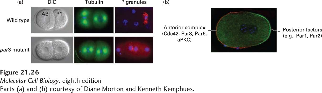
EXPERIMENTAL FIGURE 21- 26 Par proteins are asymmetrically localized in the one- cell worm embryo. (a) DIC images of wild- type and par3 mutant embryos. Notice that in wild- type cells, the AB cell is larger than the P1 cell, whereas they are the same size in the par3 mutant. The par3 mutant also has a defect in spindle orientation (as seen by microtubule staining in green) and P- granule (red) segregation. DNA is stained blue. (b) Complementary localization of the anterior Par complex (Cdc42- Par3- Par6- aPKC) (red) and posterior determinants (green) in the one- cell embryo.
[Parts (a) and (b) courtesy of Diane Morton and Kenneth Kemphues.]
[Leave] [Close]