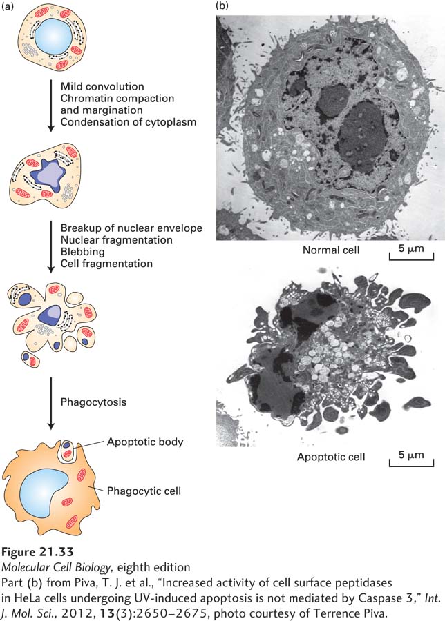
FIGURE 21- 33 Ultrastructural features of cell death by apoptosis. (a) Schematic drawings illustrating the progression of morphological changes observed in apoptotic cells. Early in apoptosis, dense chromosome condensation occurs along the nuclear periphery. The cell body also shrinks, although most organelles remain intact. Later, both the nucleus and the cytoplasm fragment, forming apoptotic bodies, which are phagocytosed by surrounding cells. (b) Photomicrographs comparing a normal cell and an apoptotic cell. Clearly visible in the latter are dense spheres of compacted chromatin as the nucleus begins to fragment.
[Part (b) from Piva, T. J. et al., “Increased activity of cell surface peptidases in HeLa cells undergoing UV- induced apoptosis is not mediated by Caspase 3,” Int. J. Mol. Sci., 2012, 13(3):2650– 2675, photo courtesy of Terrence Piva.]
[Leave] [Close]