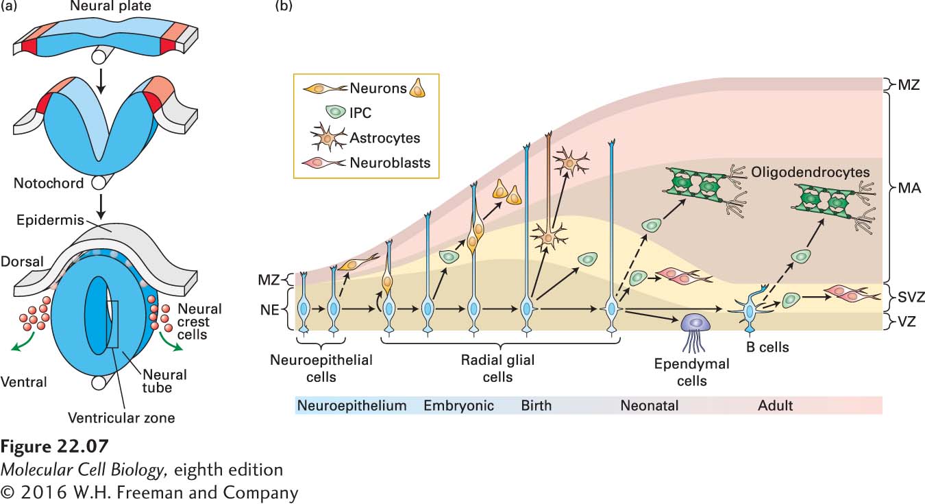
FIGURE 22- 7 Formation of the neural tube and division of neural stem cells. (a) Early in vertebrate development a part of the ectoderm rolls up and separates from the rest of the cells. This forms the epidermis (gray) and the neural tube (blue). Near the interface between the two, neural crest cells form and then migrate to contribute to skin pigmentation, nerve formation, craniofacial skeleton, heart valves, peripheral neurons, and other structures. The notochord, a rod of mesoderm for which chordates are named, provides signals that affect cell fates in the neural tube. The interior of the neural tube will become a fluid- filled series of chambers called ventricles. Neural stem cells located adjacent to the ventricles, described as being in the ventricular zone (VZ), will divide to form neurons that migrate radially outward to form the layers of the nervous system. (b) Early in development, in the neuroepithelium (NE), neuroepithelial cells divide symmetrically to generate more neuroepithelial cells. Some are also thought to generate early neurons. As development progresses and the brain epithelium thickens, neuroepithelial cells convert into radial glial cells (RGCs). RGCs divide symmetrically or asymmetrically to generate neurons or intermediate progenitor cells (IPCs), which in turn generate neurons. RGCs continue to elongate, and send an apical process down to the VZ and a basal process up to contact the meninges. Near the end of embryonic development, RGCs detach from the NE and convert into astrocytes, and also generate oligodendrocytes from IPCs. After birth, in neonates, RGCs continue to divide into neurons and oligodendrocytes, through IPCs. Others convert into ependymal cells or into adult SVZ astrocytes called type B cells that function as neural stem or progenitor cells in the subventricular zone (SVZ) of the adult brain. IPC, intermediate progenitor cell; MA, mantle; MZ, marginal zone; NE, neuroepithelium; RG, radial glia; SVZ, subventricular zone; VZ, ventricular zone. See A. Kriegstein and A. Alvarez- Buylla, 2009, Annu. Rev Neurosci. 32:149– 184.
[Leave] [Close]