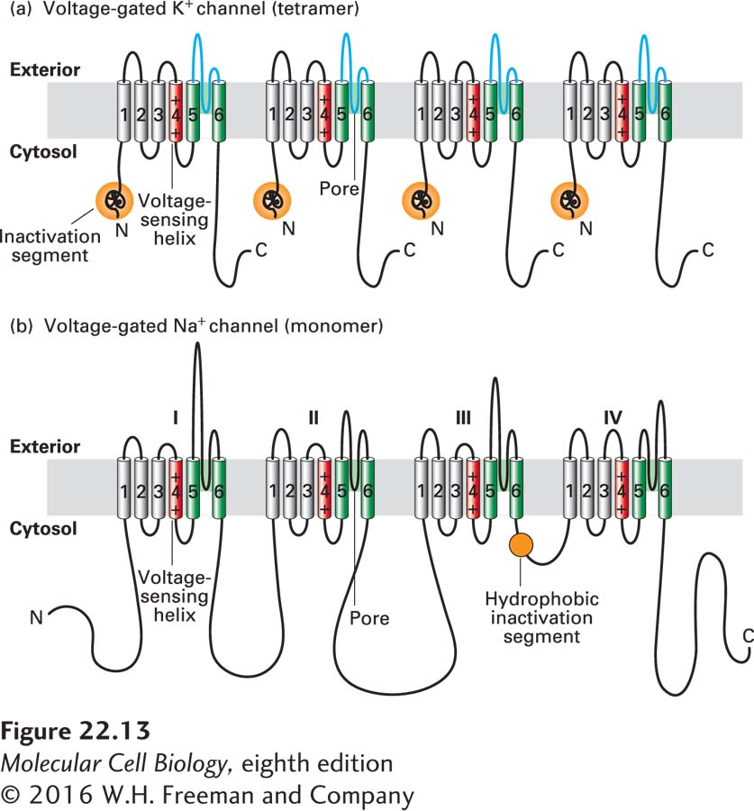
FIGURE 22- 13 Schematic depictions of the secondary structures of voltage- gated K + and Na+ channels. (a) Voltage- gated K+ channels are composed of four identical subunits, each containing 600– 700 amino acids, and six membrane- spanning α helices, S1– S6. The N- terminus of each subunit, located in the cytosol and labeled N, forms a globular domain (orange ball) essential for inactivation of the open channel. The S5 and S6 helices (green) and the P segment (blue) are homologous to those in nongated resting K+ channels, but each subunit contains four additional transmembrane α helices. One of these, S4 (red), is the primary voltage- sensing α helix and is assisted in this role by forming a stable complex with helices S1– S3. See C. Miller, 1992, Curr. Biol. 2:573, and H. Larsson et al., 1996, Neuron 16:387. (b) Voltage- gated Na+ channels are monomers containing 1800– 2000 amino acids organized into four transmembrane domains (I– IV) that are similar to the subunits in voltage- gated K+ channels. The single hydrophobic channel- inactivating segment (orange ball) is located in the cytosol between domains III and IV. Voltage- gated Ca2+ channels have a similar overall structure. Most voltage- gated ion channels also contain regulatory (β) subunits, which are not depicted here. See W. A. Catterall, 2001, Nature 409:988.
[Leave] [Close]