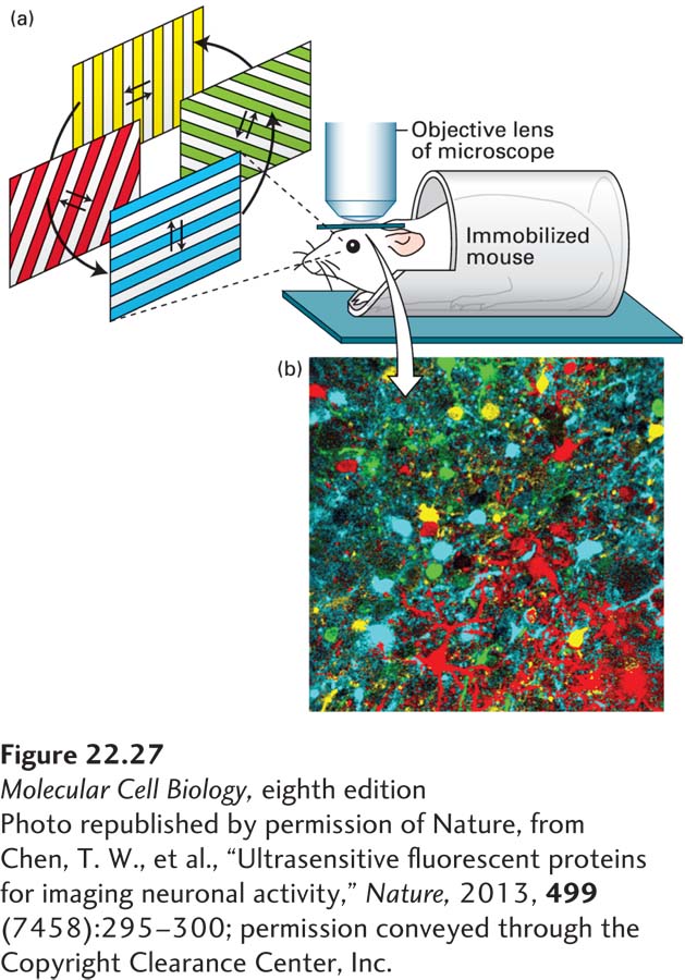
FIGURE 22- 27 Calcium indicators allow visualization of activity in neural circuits. A genetically encoded calcium indicator was expressed in neurons in the mouse visual cortex. (a) A window was made in the skull of the mouse, and a microscope (indicated by the objective lens) was used to visualize calcium transients in populations of neurons in the visual cortex while the mouse was looking at gratings that moved in different directions. Individual neurons within the visual cortex respond to specific orientations of the gratings, as detected by elevations in calcium that are visualized as increases in the fluorescence of the calcium indicator. (b) Neurons were color coded according to the orientation that elicited increases in calcium (as shown below the photo). The neurons shown in yellow respond to horizontally moving gratings, and the neurons shown in cyan respond to vertically moving gratings, while the neurons shown in green and red respond to diagonally oriented gratings. This type of experiment reveals that individual neurons are tuned to specific orientations of visual stimuli.
[Photo republished by permission of Nature, from Chen, T. W., et al., “Ultrasensitive fluorescent proteins for imaging neuronal activity,” Nature, 2013, 499(7458):295– 300; permission conveyed through the Copyright Clearance Center, Inc.]
[Leave] [Close]