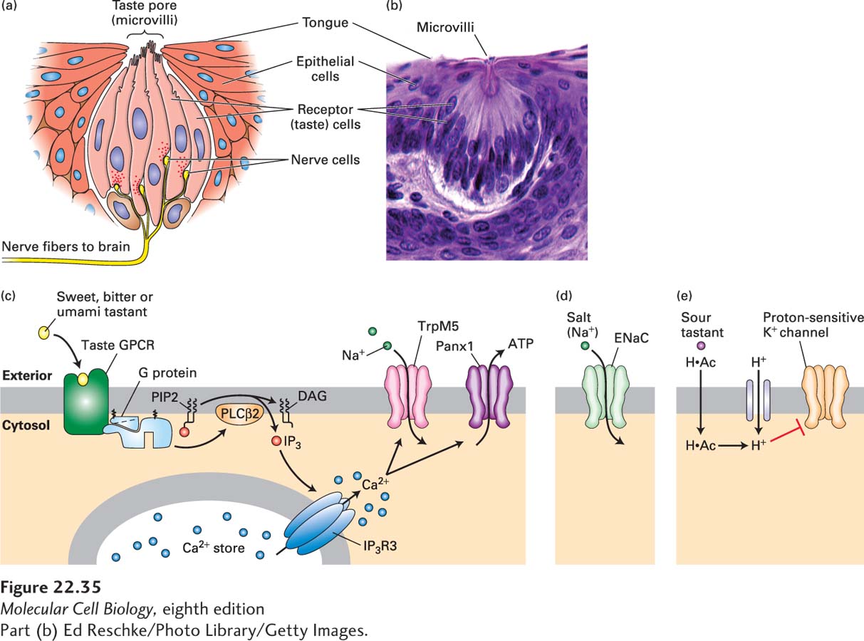
FIGURE 22- 35 The sense of taste. (a) The taste cells (pink) in a mammalian taste bud contact the nerve cells (yellow). The chemical signals arrive at the microvilli seen at the top. (b) Micrograph of a mammalian taste bud, showing the receptor cells. The microvilli are barely visible at the top of the taste bud, indicated by the label. (c) Sweet, bitter, and umami ligands bind specific taste GPCRs expressed in Type II receptor cells, activating a phosphoinositide pathway that elevates cytosolic Ca2+ · Ca2+ in turn binds to and opens a Ca2+-gated Na+ channel, TrpM5, leading to an influx of Na+ and membrane depolarization. The combined action of elevated Ca2+ and membrane depolarization opens the large pores of an unusual membrane channel termed Panx1, resulting in release of ATP and probably other signaling molecules into the extracellular space. ATP and these other molecules stimulate the nerve cells that will ultimately carry the information to the brain. (d) Salt is detected by direct permeation of Na+ ions through membrane ion channels, including the ENaC channel, directly depolarizing the plasma membrane. (e) Organic acids like acetic acid diffuse in their protonated form (H·Ac) through the plasma membrane and dissociate into an anion and proton, acidifying the cytosol. Entry of strong acids like HCl is facilitated by a proton channel in the apical membrane of the sour- sensing cells that enables protons to reach the cytosol. Intracellular H+ is believed to block a proton- sensitive K+ channel (as yet unidentified) and thus depolarize the membrane. Voltage- gated Ca2+ channels would open, leading to an elevation in cytosolic Ca2+ that triggers exocytosis of synaptic vesicles that are not depicted. See N. Chaudhari and S. D. Roper, 2010, J. Cell Biol. 190:285; and S. Frings, 2010, PNAS 107:21955.
[Part (b) Ed Reschke/Photo Library/Getty Images.]
[Leave] [Close]