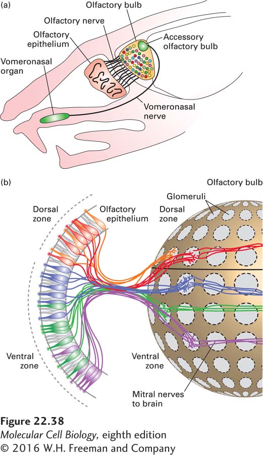
FIGURE 22- 38 The anatomy of olfaction in the mouse. (a) Schematic representation of a sagittal section through an adult mouse head. Axons of the olfactory receptor neurons (ORNs) in the main olfactory epithelium bundle to form the olfactory nerve and innervate the olfactory bulb. Each ORN of the main olfactory epithelium expresses only one odorant receptor gene. The vomeronasal organ and the accessory olfactory bulb are involved in pheromone sensing. (b) All of the olfactory receptor neurons that express a single type of receptor send their axons to the same glomerulus. In this figure each color represents the neural connections for each distinct expressed receptor. The glomeruli are located in the olfactory bulb near the brain; in the glomeruli, the ORNs synapse with mitral neurons; each mitral neuron has its dendrites localized to a single glomerulus and its corresponding ORNs, thus carrying information about a particular odorant to higher centers of the brain. Each glomerulus thus receives innervation from sensory neurons expressing a single odorant receptor, providing the anatomical basis of the olfactory sensory map. See T. Komiyama and l. Luo, 2005, Curr. Opin. Neurobiol. 16:67– 73 and S. Demaria and J. Ngai, 2010, J. Cell Biol. 191:443.
[Leave] [Close]