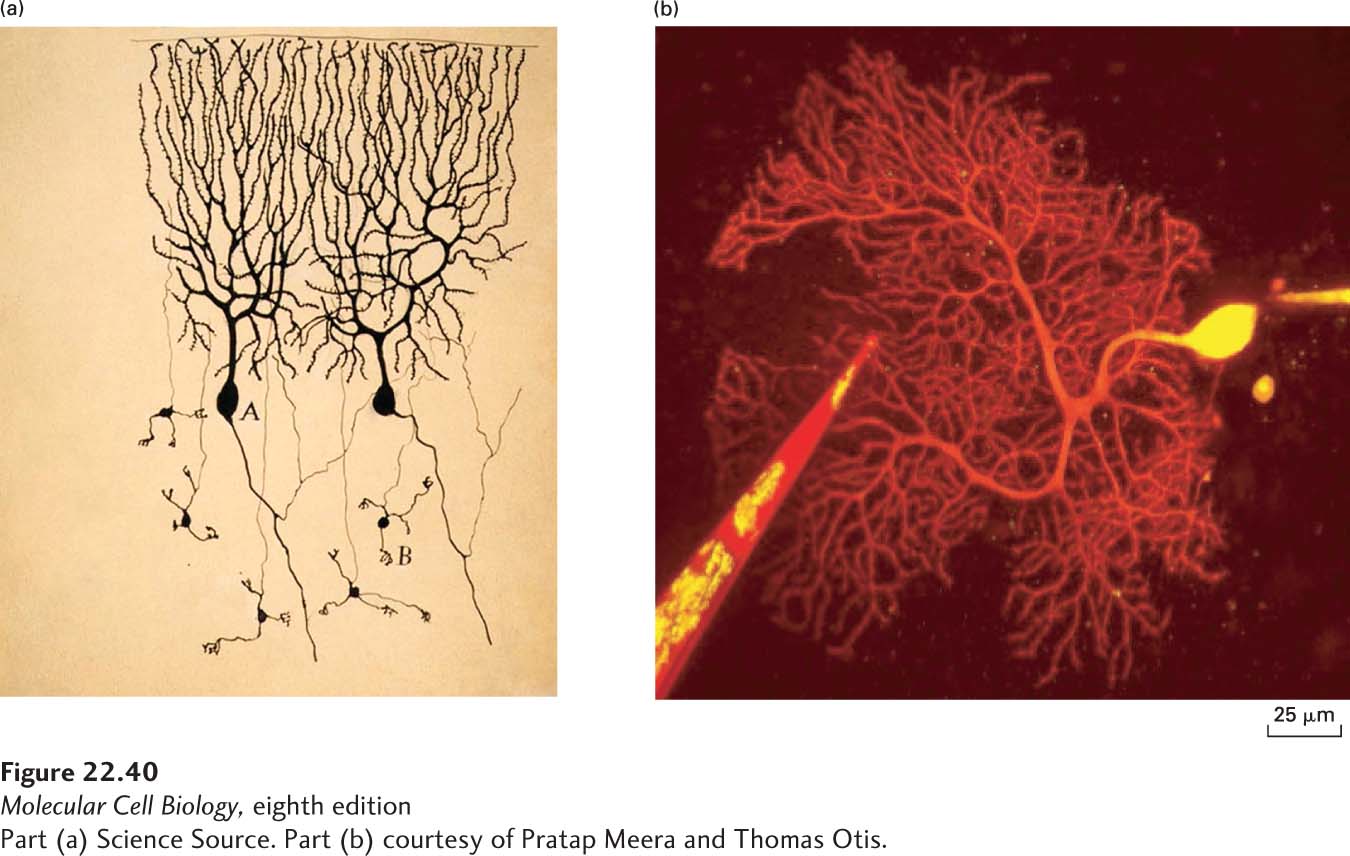
FIGURE 22- 40 Visualizing dendritic spines. (a) Santiago Ramón y Cajal used the Golgi staining method to visualize individual neurons in the cerebellum of a pigeon in 1899. This method permitted Ramón y Cajal to visualize individual neurons in the brain; the tissue is densely packed with neurons but the Golgi stain only labels sparse neurons in the tissue. Using this approach, he argued that the brain was composed of individual neurons that communicated with each other at sites of contact. The postsynaptic compartment of excitatory synapses consists of a spiny protuberance from the dendrite, called a spine. Ramón y Cajal detected these spines in neurons (here in the Purkinje neurons of the cerebellum) and hypothesized that memories could be stored as changes in the number and shape of the spines. In modern- day approaches, fluorescent proteins can be delivered using a microelectrode or expressed genetically to allow visualization of a single neuron in tissue. (b) A red fluorescent dye is delivered to a single Purkinje neuron in mouse cerebellum by a microelectrode and is visualized by two- photon microscopy. At higher resolution, one can image spine dynamics using time- lapse microscopy, and in this way directly demonstrate changes in synaptic connectivity with experience. In this image, a second electrode filled with a red fluorescent dye is used to stimulate synapses forming onto the labeled neurons.
[Part (a) Science Source. Part (b) courtesy of Pratap Meera and Thomas Otis.]
[Leave] [Close]