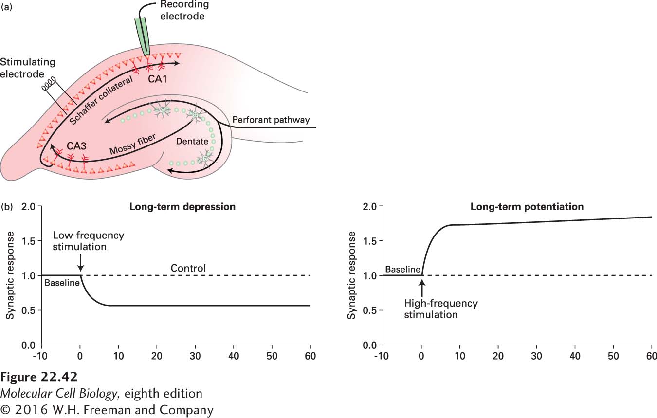
FIGURE 22- 42 Synaptic plasticity in the mouse hippocampus: long- term potentiation (LTP) and long- term depression (LTD). (a) The mouse hippocampus can be dissected from mouse brain cut into transverse slices, preserving the three sequential synaptic pathways. In the perforant pathway, axons from the entorhinal cortex project to form synapses on dendrites of dentate granule cells (green circles); in the mossy fiber pathway, dentate granule axons synapse on CA3 pyramidal neuron (red triangles) dendrites; and in the Schaffer collateral pathway, CA3 axons synapse on CA1 pyramidal neuron (red triangles) dendrites. The dentate granule cells (green) and the CA3 and CA1 pyramidal cell bodies (red) form discrete somatic layers projecting axons and dendrites into defined pathways. Electrodes can be used to stimulate axonal afferents and record from postsynaptic follower cells, as illustrated for the Schaffer collateral (CA3- CA1) pathway. (b) Trains of low- frequency stimulation or high- frequency stimulation to the axonal fibers produce sustained decreases or increases in synaptic strength, which are measured as the postsynaptic response to a test stimulus. These forms of plasticity are known as long- term depression (LTD) and long- term potentiation (LTP). See V. M. Ho, J. A. Lee, and K. C. Martin, 2011, Science 334:623– 628.
[Leave] [Close]