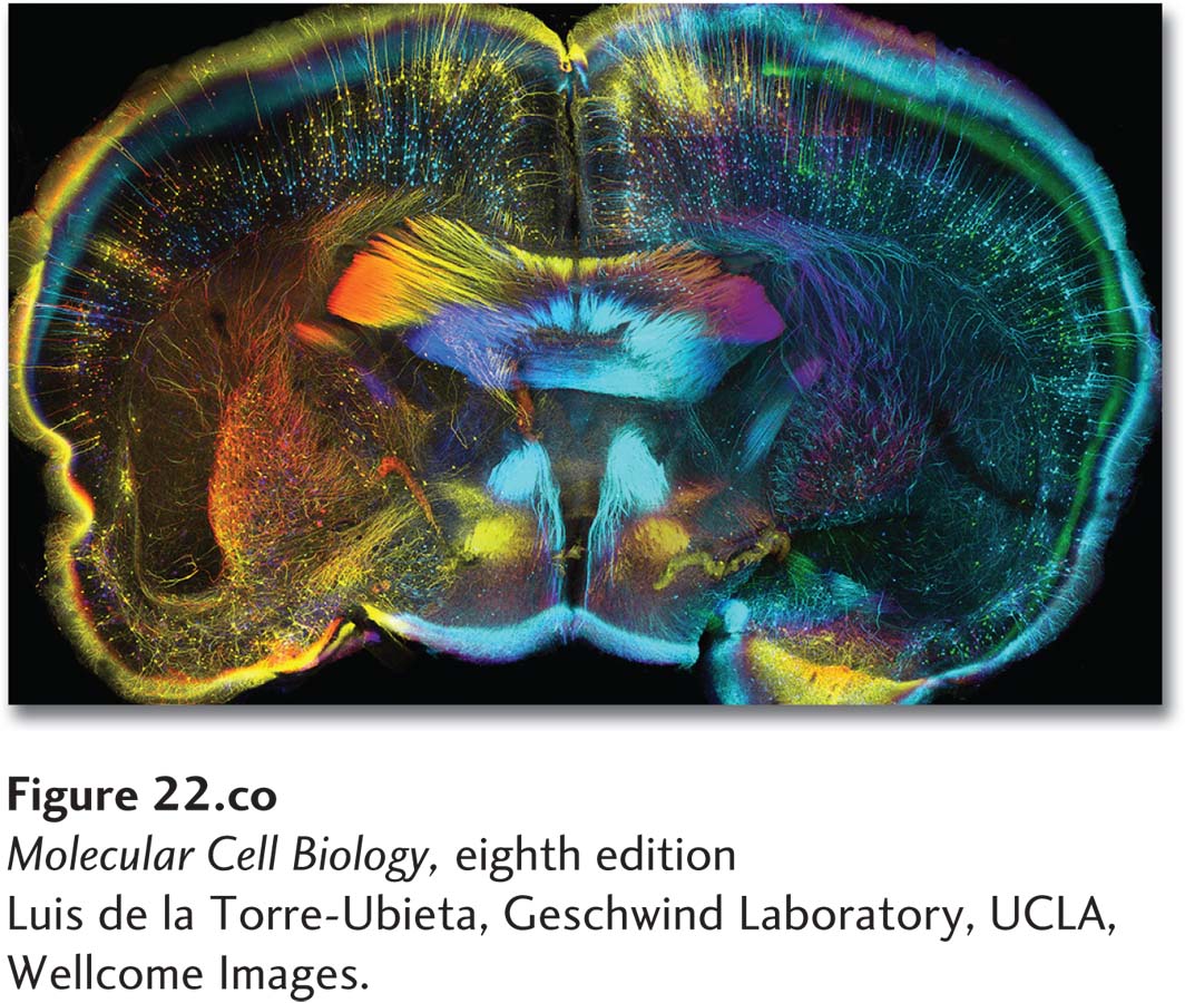
Coronal slice of CLARITY- treated adult mouse brain expressing GFP in a subset of neurons (Thy1- GFP). CLARITY renders tissue optically transparent, permitting deep and complete imaging of tissues, including brains. Section was stained with antibodies to GFP and color- coded by depth to facilitate individual neuron visualization. The final image is assembled from over 8500 individual images digitally stitched together over a 750- μm thick piece of brain. This approach provides unprecedented opportunity to image intact brains at cellular resolution, paving the way to a comprehensive understanding of how the brain is wired.
[Luis de la Torre- Ubieta, Geschwind Laboratory, UCLA, Wellcome Images.]
[Leave] [Close]