image
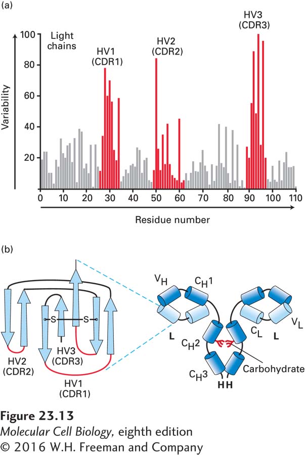
FIGURE 23-13 Hypervariable regions and the immunoglobulin fold. (a) Amino acid variability varies with residue position in Ig light chains. Here the percentage of variable-region sequences with variant amino acids is plotted for each position in the sequence. Positions for which many different amino acid side chains are present are assigned a high variability index; those that are invariant among the sequences compared are assigned a value of 0. This analysis reveals three regions of increased variability: hypervariability (HV) regions 1, 2, and 3; these regions are also called complementarity-determining regions (CDRs). (b) Volume-rendered depiction of F(ab′)2 fragment (right) and ribbon diagram of a typical Ig light-chain variable region (VL) with the positions of the hypervariable regions indicated in red (left). The hypervariable regions are found in the loops that connect the β strands and make contact with antigen. The β strands (rendered as arrows) make up two β sheets and constitute the framework region. Each variable and constant domain has this characteristic three-dimensional structure, called the immunoglobulin fold. L = light chain; H = heavy chain; VH = heavy-chain variable region; VL = light-chain variable region; CH1, CH2, CH3 = heavy-chain constant domains; CL = light-chain constant region.