image
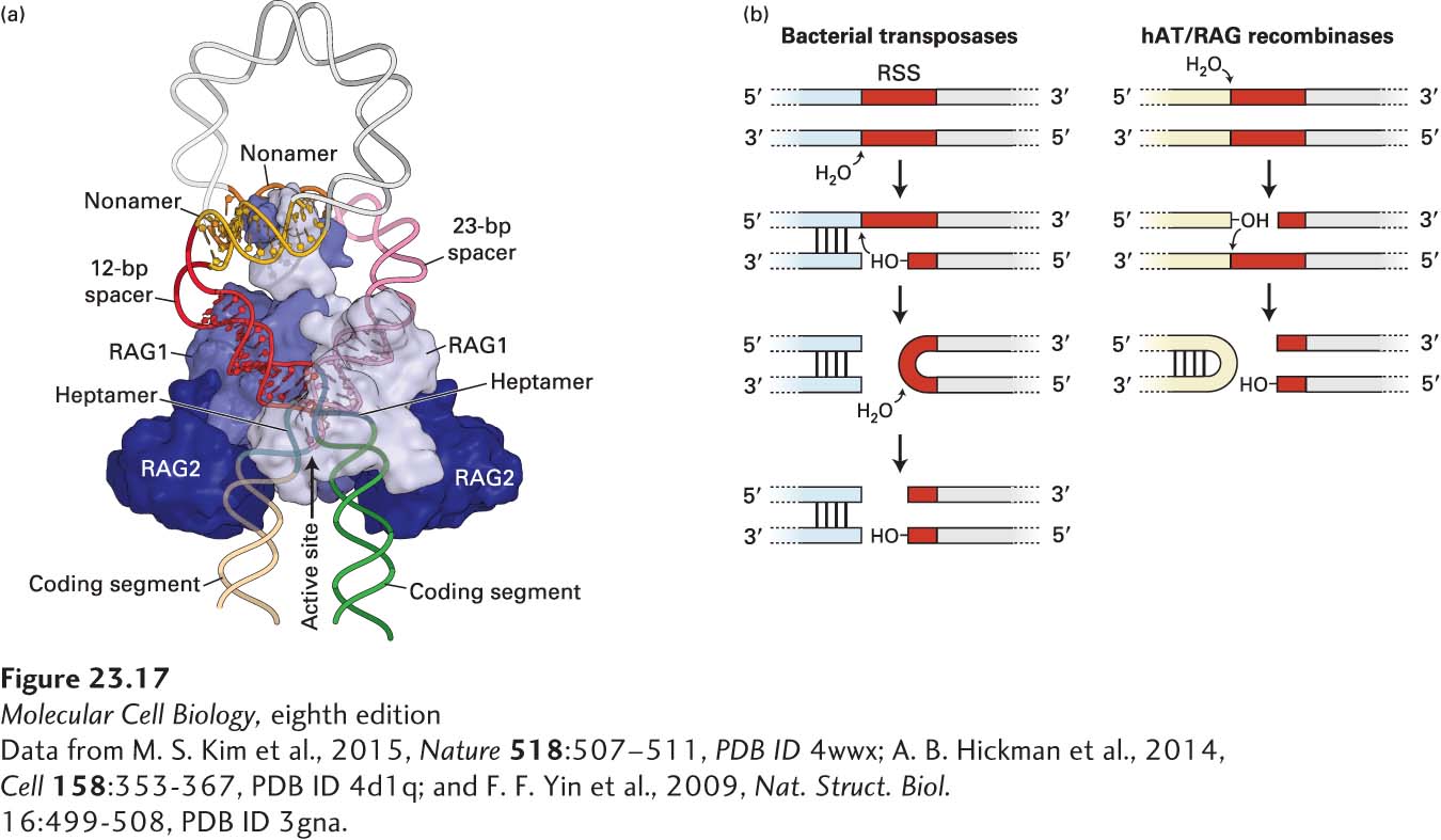
FIGURE 23-17 RAG1/RAG2 structure. (a) RAG1/RAG2 is shown in complex with the recombination signal sequences, positioning the 12- and 23-bp spacer sequences to enable cleavage at the boundary of the coding sequence and the heptamer of the RSS. (b) DNA can be cleaved by hairpin-forming bacterial and eukaryotic transposases, the evolutionary precursors of the RAG1/RAG2 complex. Shown here is the generation of a single-strand break, followed by an attack by the newly generated 3′ hydroxyl on the complementary strand to form a hairpin and a double-strand break.
[Data from M. S. Kim et al., 2015, Nature 518:507–511, PDB ID 4wwx; A. B. Hickman et al., 2014, Cell 158:353-367, PDB ID 4d1q; and F. F. Yin et al., 2009, Nat. Struct. Biol. 16:499-508, PDB ID 3gna.]