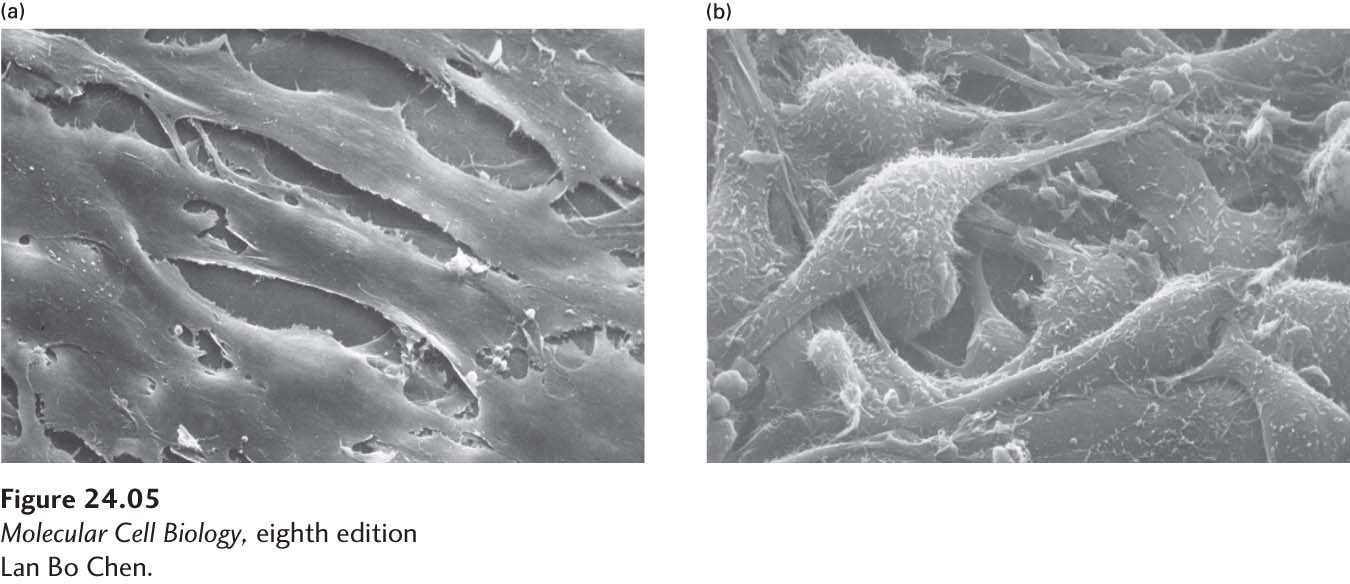
EXPERIMENTAL FIGURE 24- 5 Scanning electron micrographs reveal the organizational and morphological differences between normal and transformed 3T3 cells. (a) Cultured mouse fibroblasts called 3T3 cells are normally elongated and are aligned and closely packed in an orderly fashion. (b) 3T3 cells transformed by an oncogene encoded by Rous sarcoma virus are rounded and covered with small hairlike processes and bulbous projections. The transformed cells have lost the side- by- side organization of the normal cells and grow one atop the other. These transformed cells have many of the same properties as malignant cells. Similar changes are seen in cells transfected with DNA from human cancers containing the rasD oncogene.
[Lan Bo Chen.]
[Leave] [Close]