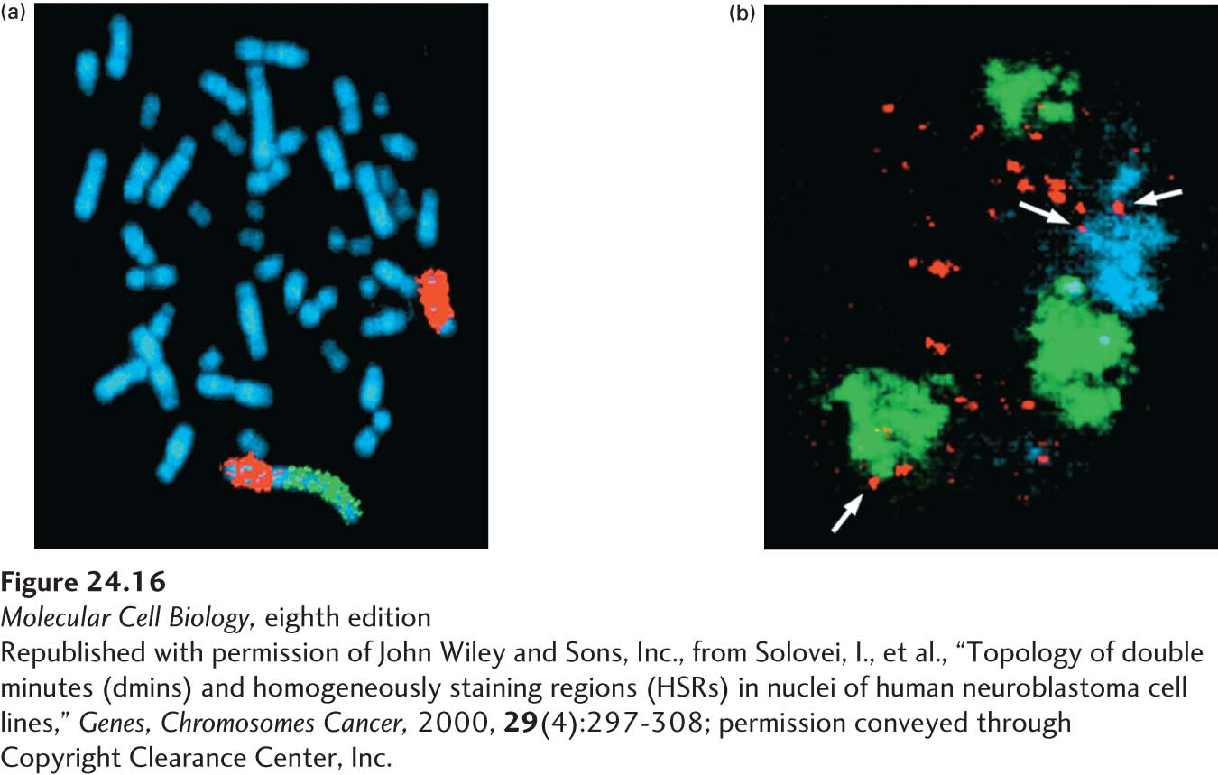
EXPERIMENTAL FIGURE 24- 16 DNA amplifications in stained chromosomes take two forms, both visible under the light microscope. (a) Homogeneously staining regions (HSRs) in a human chromosome from a neuroblastoma cell. The chromosomes are uniformly stained with a blue dye so that all can be seen. Specific DNA sequences were detected using fluorescent in situ hybridization (FISH), in which fluorescently labeled DNA clones are hybridized to denatured DNA in the chromosomes. The chromosome 4 pair is marked by in situ hybridization with a chromosome paint probe for the long arm of chromosome 4 (red). On one of the chromosome 4’s an HSR is visible after hybridizing with a probe for the N- MYC gene (green), which is amplified in this neuroblastoma cell. (b) Optical sections through nucleus from a human neuroblastoma cell that contain so- called double- minute chromosomes. The normal chromosomes are the green and blue structures; the double- minute chromosomes are the many small red dots. Arrows indicate double minutes associated with the surface or interior of the normal chromosomes.
[Republished with permission of John Wiley and Sons, Inc., from Solovei, I., et al., “Topology of double minutes (dmins) and homogeneously staining regions (HSRs) in nuclei of human neuroblastoma cell lines, “Genes, Chromosomes Cancer, 2000, 29(4):297- 308; permission conveyed through Copyright Clearance Center, Inc.]
[Leave] [Close]