image
FIGURE 3-23 Active site of the enzyme trypsin. (a) An enzyme’s active site (outlined by dashed line) is composed of a substrate-binding site (blue), which binds specifically to a substrate, and a catalytic site (purple), which carries out catalysis. (b) A hybrid surface/ribbon representation of a portion of the serine protease trypsin. Clearly visible are the active-site cleft containing the catalytic site (purple, includes the key catalytic triad of Ser-195, Asp-102, and His-57, see also Figure 3-27) and a portion of the substrate-binding site called the side-chain-specificity binding pocket (blue).
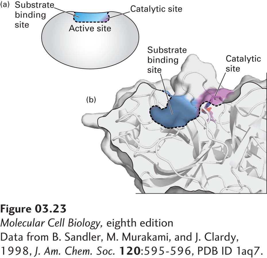
[Data from B. Sandler, M. Murakami, and J. Clardy, 1998, J. Am. Chem. Soc. 120:595-596, PDB ID 1aq7.]