image
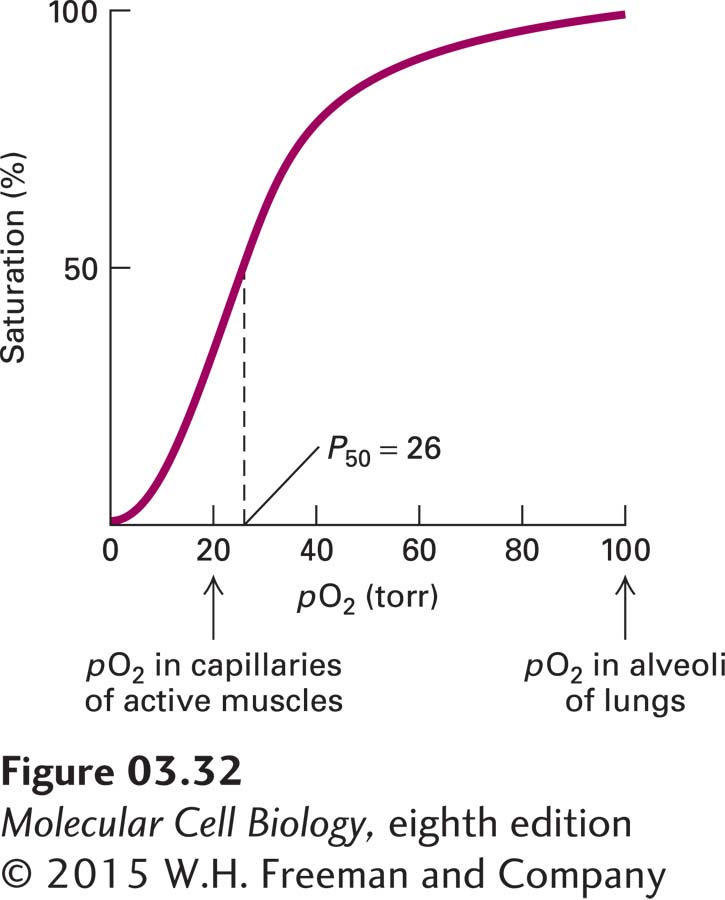
EXPERIMENTAL FIGURE 3-32 Hemoglobin binds oxygen cooperatively. Each tetrameric hemoglobin molecule has four oxygen-binding sites; at saturation, all the sites are loaded with oxygen. The oxygen concentration in tissues is commonly measured as the partial pressure of oxygen (pO2) in torr units (a standard measure of pressure equivalent to 1 mm of mercury under standard conditions). P50 is the pO2 at which half the oxygen-binding sites are occupied; it is somewhat analogous to the Km for an enzymatic reaction. The large change in the amount of oxygen bound over a small range of pO2 values permits efficient unloading of oxygen in peripheral tissues such as muscle. The sigmoidal shape of a plot of saturation versus ligand concentration is indicative of cooperative binding, in which the binding of one oxygen molecule allosterically influences the binding of subsequent oxygens. In the absence of cooperative binding, a binding curve is a hyperbola, similar to the curves in Figure 3-24. See J. M. Berg et al., 2015, Biochemistry, 8th ed., Macmillan.