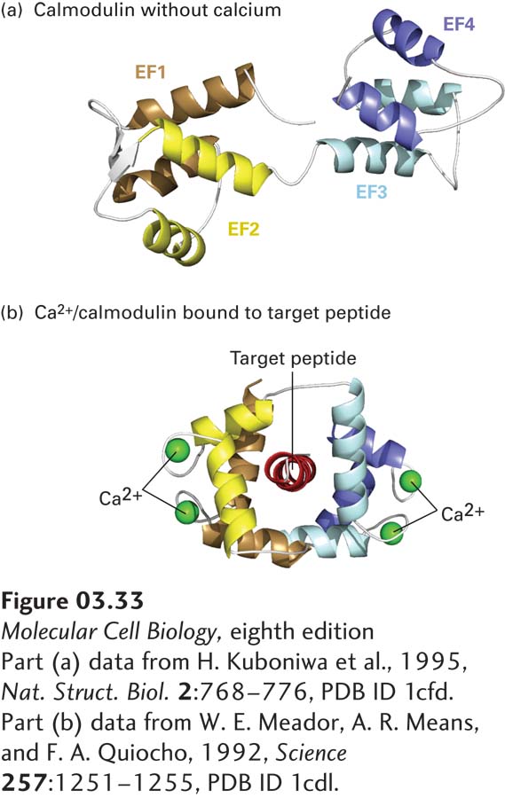
image
FIGURE 3-33 Conformational changes induced by Ca2+ binding to calmodulin. Calmodulin is a widely distributed cytosolic protein that contains four Ca2+-binding sites, one in each of its EF hand (helix-loop-helix) motifs (EF1-ER4, see also Figure 3-10). At cytosolic Ca2+ concentrations above about 5 × 10⁻⁷ M, binding of Ca2+ to calmodulin changes the protein’s conformation from the dumbbell-shaped, Ca2+-free form (a) to one in which hydrophobic side chains become more exposed to solvent. The resulting Ca2+/calmodulin complex can wrap around exposed helices (target peptides) with specialized sequences in various target proteins (b), thereby altering their activities.
[Part (a) data from H. Kuboniwa et al., 1995, Nat. Struct. Biol. 2:768–776, PDB ID 1cfd. Part (b) data from W. E. Meador, A. R. Means, and F. A. Quiocho, 1992, Science 257:1251–1255, PDB ID 1cdl.]