image
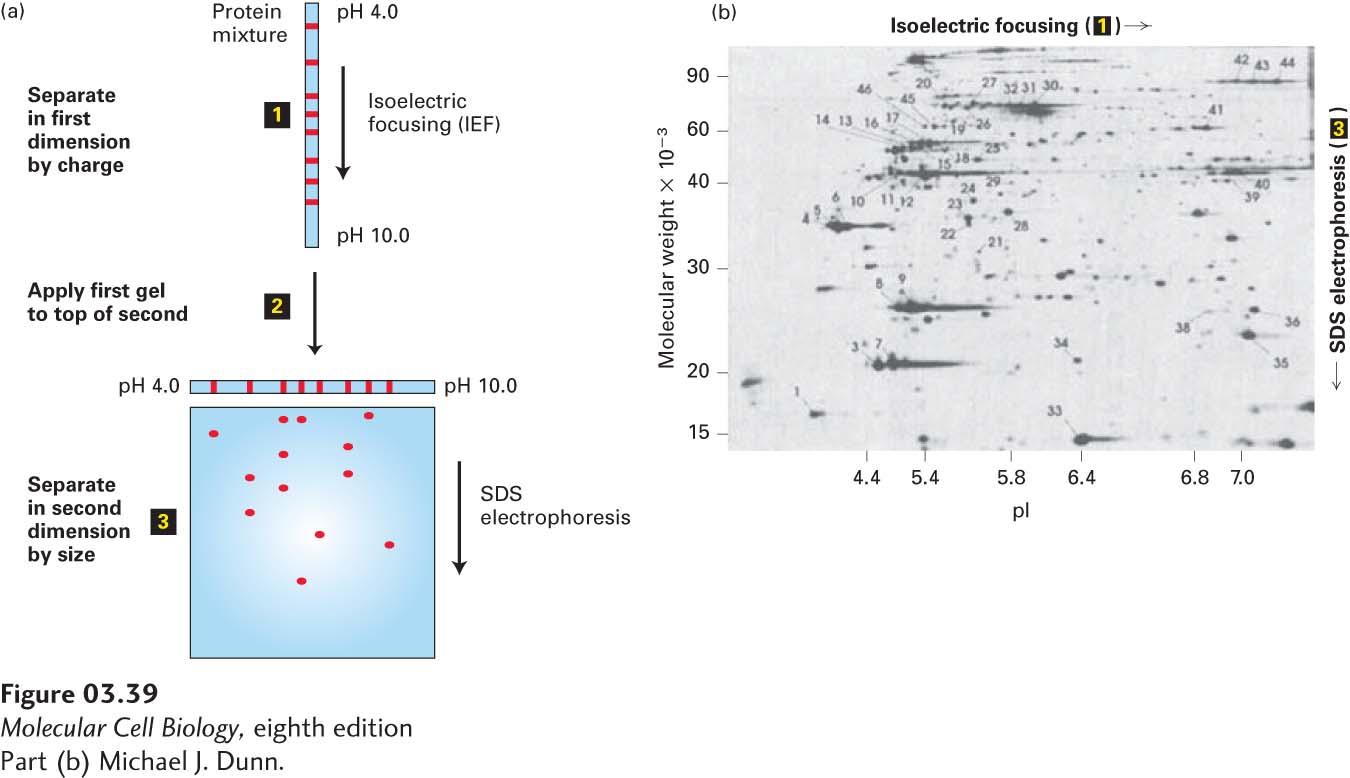
EXPERIMENTAL FIGURE 3-39 Two-dimensional gel electrophoresis separates proteins on the basis of charge and mass. (a) In this technique, proteins are first separated into bands on the basis of their charges by isoelectric focusing (step 1). The resulting gel strip is applied to an SDS-polyacrylamide gel (step 2), and the proteins are separated into spots by mass (step 3). (b) In this two-dimensional electrophoresis gel of a protein extract from cultured cells, each spot represents a single polypeptide. Polypeptides can be detected by dyes, as here, or by other techniques, such as autoradiography. Each polypeptide is characterized by its isoelectric point (pI) and molecular weight.
[Part (b) Michael J. Dunn.]