image
EXPERIMENTAL FIGURE 3-41 Immunoblotting (IP, or Western blotting) and co-immunoprecipitation (co-IP) can detect specific proteins and their binding partners. (a) Immunoblotting method. Step 1: After a protein mixture has been electrophoresed through an SDS gel, the separated bands (or spots, for two-dimensional gel electrophoresis) are transferred (blotted) from the gel onto a porous membrane from which the protein is not readily removed. Individual proteins (represented by blue ovals) are not visible at this stage. Step 2: The membrane is flooded with a solution of an antibody (Ab1) specific for the protein of interest and allowed to incubate for a while. Ab1 binds to the protein of interest (second from the top), but not to any other proteins attached to the membrane, forming a layer of antibody molecules coincident with the protein (whose position still cannot be seen at this point). Then the membrane is washed to remove unbound Ab1. Step 3: The membrane is incubated with a second antibody (Ab2) that specifically recognizes and binds to the first (Ab1). This second antibody is covalently linked to an enzyme that catalyzes a chromogenic reaction or releases light (e.g., chemiluminescence), a radioactive isotope, or some other substance whose presence can be detected with great sensitivity. Step 4: Finally, the location and amount of bound Ab2 are detected (e.g., by its color for a chromogenic reaction or by detectors or film that measure the light released by chemiluminescence), permitting the electrophoretic mobility (and therefore the mass) of the protein of interest to be determined as well as its quantity (based on band intensity). (b) Immunoblotting was used to detect intracellular receptors and the influence of exposure to a ligand for one of the receptors. In this experiment, cells that are precursors to red blood cells were maintained in vitro in petri dishes and then treated with no ligand (−, leftmost and rightmost lanes) or a ligand that binds to GR, the glucocorticoid receptor (+, center lane). The cells were then lysed in detergent, and immunoblotting (Western blotting) was performed on the total cell lysates using three different antibodies that bind to GR (anti-GR), to a receptor called PPARα (anti-PPARα), or to an abundant intracellular protein, actin, whose presence and abundance was not expected to be sensitive to treatment with the ligand. The equal intensities of the immunoblotting bands detected using the anti-actin antibody (bottom box) provided a “loading control,” which established that essentially equal amounts of cell lysate were applied (loaded) in each lane of the gel. The approximately equal intensities of the bands for both GR and PPARα with or without prior incubation of the cells with the GR ligand showed that the ligand did not substantially alter the amounts of either of these proteins in the cells. Portions of the same cell lysates used for the immunoblotting in part (b) were also used for the immunoprecipitation/immunoblotting shown in part (c). (c) Immunoprecipitation (IP) followed by immunoblotting (together called co-IP) was used to determine if the GR ligand can induce formation of a stable complex that contains both GR and PPARα. Portions of the cell lysates were immunoprecipitated with an antibody to GR (left and center lanes) or a control antibody (right lane) that cannot bind to either GR or PPARα. The immunoprecipitates were separated from the rest of the lysates by centrifugation and then analyzed by immunoblotting with either anti-GR (top box) or anti-PPARα (bottom box). As expected, the top box shows that the GR protein was detected in the immunoprecipitates generated using the α-GR when the same anti-GR antibody was used for the immunoblotting, but not in the immunoprecipitates generated with the control antibody (no band observed). Strikingly, when one examines the immunoprecipitates by immunoblotting with the anti-PPARα antibody (bottom box), a substantial amount of PPARα is seen when the GR ligand is present (center lane), whereas little co-precipitates in the absence of the GR ligand (left lane) or in the control immunoprecipitate (right lane). These results indicate that the GR ligand induces formation of a complex containing both the glucocorticoid receptor and the PPARα proteins. These results do not establish whether or not the GR and PPARα proteins bind directly to each other when the GR ligand is present or if there are additional molecules in the complex that act as intermediates holding the GR and PPARα tightly together when the ligand is present.
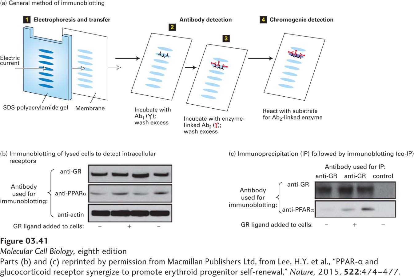
[Parts (b) and (c) reprinted by permission from Macmillan Publishers Ltd, from Lee, H.Y. et al., “PPAR-α and glucocorticoid receptor synergize to promote erythroid progenitor self-renewal,” Nature, 2015, 522:474–477.]