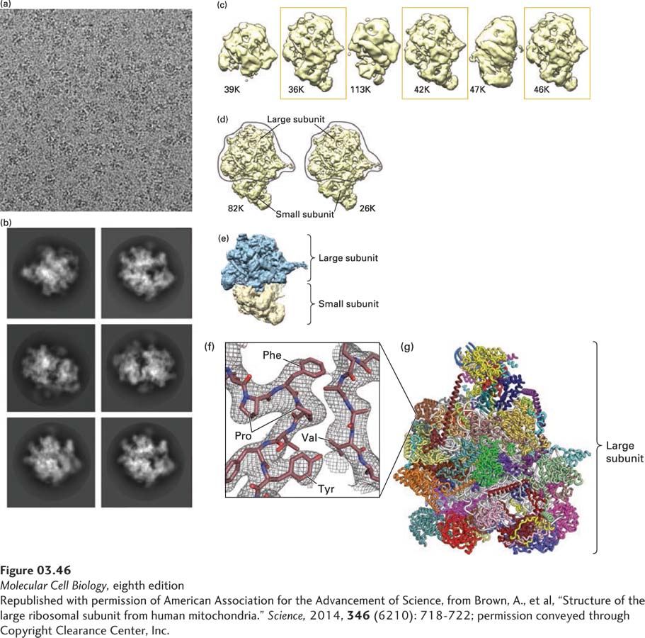
EXPERIMENTAL FIGURE 3- 46 Cryoelectron microscopy analysis of the structure of the human mitochondrial ribosome. The mitochondrion is a complex, multifunctional intracellular organelle best known for its ability to synthesize the energy carrier ATP (see Chapter 12). Human mitochondria can synthesize proteins encoded by mitochondrial DNA using large (1.7 MDa), multi- protein (at least 78) and multi- RNA complexes called mitochondrial ribosomes that differ somewhat from cytoplasmic ribosomes. (a) Cryoelectron micrograph of isolated human mitochondrial ribosomes. The low contrast between the ribosomes and the buffer solution makes it difficult to clearly see individual, frozen ribosome particles, which are oriented randomly in the image. (b) Automated image processing of 323,292 individual particles permits their grouping into classes based on orientation and averaging of the images within each class to generate clearer images of the ribosome. (c) Additional computational analysis generates distinct structures, each based on tens of thousands of individual particles (the number of particles analyzed for each structure in thousands [K] is shown beneath each). The structures enclosed in boxes were selected for additional analysis, which produced the two very similar models shown in (d) containing virtually identical large subunits. (e) Color- coded, low- resolution model of the electron density of the large (blue) and small (yellow) subunits. The conformational heterogeneity of the small subunit prevented its high- resolution structure determination from the data shown here. (f) High- magnification view of the experimentally determined electron density (meshwork) from a portion of one of the proteins in the large subunit illustrates how the electron density is used to build the superimposed molecular model of polypeptide chains. In this very small portion of one protein within the large subunit, the side chains of proline (Pro), phenylalanine (Phe), valine (Val), and tyrosine (Tyr) residues are easily seen and demonstrate the power of cryoelectron microscopy to determine protein structures at very high resolutions. (g) Model of the 48 protein subunits (different colors) in the large subunit determined at 3.4 Å resolution.
[Republished with permission of American Association for the Advancement of Science, from Brown, A., et al, “Structure of the large ribosomal subunit from human mitochondria.” Science, 2014, 346 (6210): 718- 722; permission conveyed through Copyright Clearance Center, Inc.]
[Leave] [Close]