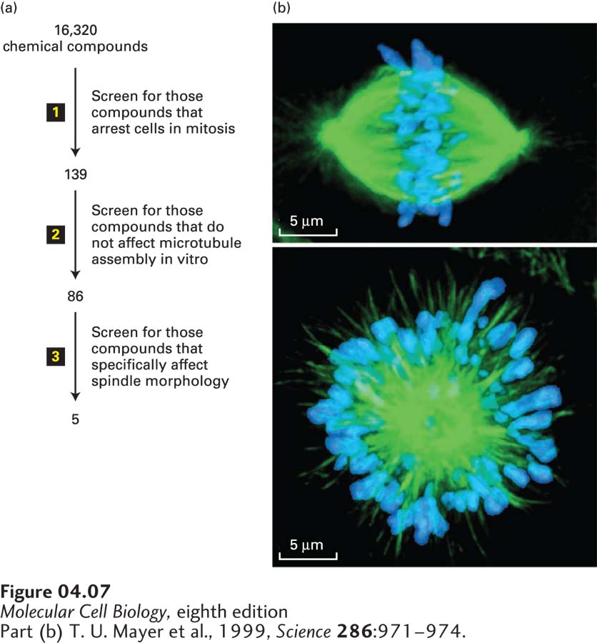
FIGURE 4- 7 Screening for drugs that affect specific biological processes. (a) In this example, a chemical library of 16,320 different chemicals was subjected to a series of screens for inhibitors of mitosis. Since such an inhibitor is expected to arrest cells at the mitotic stage of the cell cycle, the first screen (step 1) was to see if any of the chemicals enhanced the level of a marker specific for mitotic cells; this screen yielded 139 candidates. Microtubules make up the structure of the mitotic spindle, and the researchers were not interested in new drugs that target microtubules, so in the second screen (step 2) they tested the 139 compounds for their ability to affect microtubule assembly; this test eliminated 53 candidates. Immunofluorescence microscopy with antibodies to tubulin (the major subunit of microtubules), together with a stain for DNA, was then used in the third screen (step 3) to identify compounds that disrupt the structure of the spindle. (b) Localization of tubulin (green) and DNA (blue) for an untreated mitotic spindle (top) and one treated with one of the recovered compounds, now called monastrol. Monastrol inhibits a microtubule- based motor protein called kinesin- 5, discussed in Chapter 18, that is necessary to separate the poles of the mitotic spindle. When kinesin- 5 is inhibited, the two poles remain associated to give a monopolar spindle.
[Part (b) T. U. Mayer et al., 1999, Science 286:971– 974.]
[Leave] [Close]