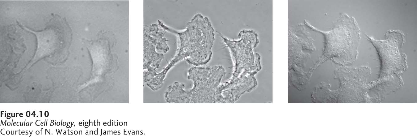
FIGURE 4- 10 Live cells can be visualized by microscopy techniques that generate contrast by interference. These micrographs show live, cultured macrophage cells viewed by bright- field microscopy (left), phase- contrast microscopy (middle), and differential- interference- contrast (DIC) microscopy (right). In a phase- contrast image, cells are surrounded by alternating dark and light bands; in- focus and out- of- focus details are simultaneously imaged in a phase- contrast microscope. In a DIC image, cells appear in pseudorelief. Because only a narrow in- focus region is imaged, a DIC image is an optical slice through the object.
[Courtesy of N. Watson and James Evans.]
[Leave] [Close]