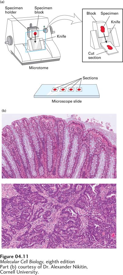
FIGURE 4- 11 Tissues for light microscopy are commonly fixed, embedded in a solid medium, and cut into thin sections. (a) A fixed tissue is dehydrated by soaking in a series of alcohol- water solutions, ending with an organic solvent compatible with the embedding medium. To embed the tissue for sectioning, the tissue is placed in liquid paraffin. After the block containing the specimen has hardened, it is mounted on the arm of a microtome, and slices are cut with a knife. Typical sections cut for light microscopy are 0.5 to 50 µm thick. The sections are collected on microscope slides and stained with an appropriate agent. (b) Sections of normal (top) and cancerous (adenocarcinoma, bottom) human colon stained with H&E stain. Notice the disorganization of the cells in the cancer tissue.
[Part (b) courtesy of Dr. Alexander Nikitin, Cornell University.]
[Leave] [Close]