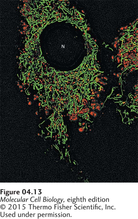
EXPERIMENTAL FIGURE 4- 13 Location of lysosomes and mitochondria in a cultured living bovine pulmonary artery endothelial cell. The cell was stained with a green- fluorescing dye that is specifically bound to mitochondria and a red- fluorescing dye that is specifically incorporated into lysosomes. The image was sharpened using a deconvolution computer program discussed later in the chapter. N, nucleus.
[© 2015 Thermo Fisher Scientific, Inc. Used under permission.]
[Leave] [Close]