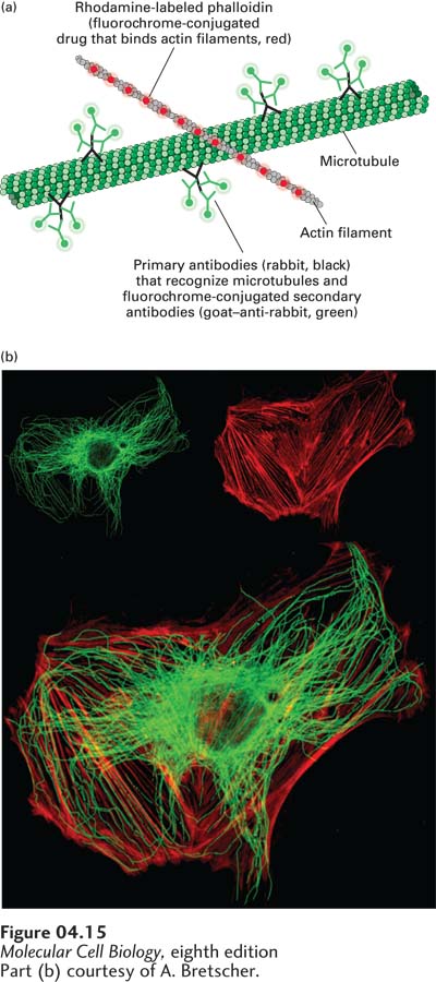
EXPERIMENTAL FIGURE 4- 15 Double- label fluorescence microscopy can visualize the relative distributions of two proteins. In double- label fluorescence microscopy, each protein must be labeled with a different fluorochrome. (a) A cultured cell was fixed and permeabilized and then incubated with Rhodamine- labeled phalloidin, a reagent that specifically binds to filamentous actin. It was also incubated with rabbit antibodies to tubulin, the major component of microtubules, followed by a fluorescein- labeled secondary goat– anti- rabbit antibody. (b) The upper panels show the fluorescein- stained tubulin (left) and Rhodamine- stained actin (right), and the lower panel shows the electronically merged images.
[Part (b) courtesy of A. Bretscher.]
[Leave] [Close]