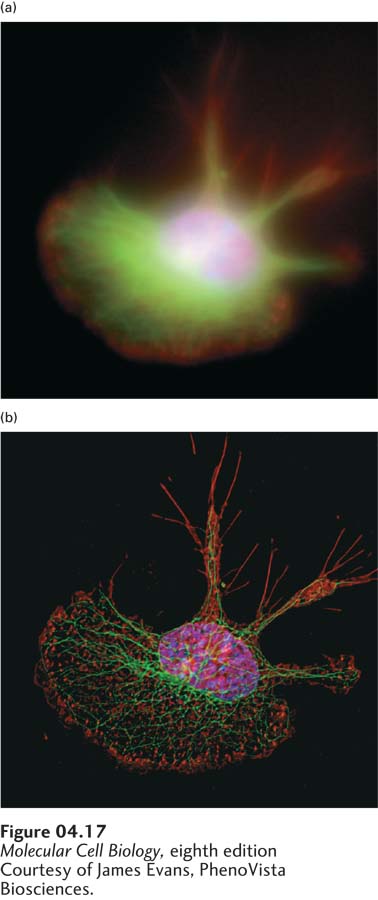
EXPERIMENTAL FIGURE 4- 17 Deconvolution fluorescence microscopy yields high- resolution optical sections that can be reconstructed to create one three- dimensional image. A macrophage cell was stained with fluorochrome- labeled reagents specific for DNA (blue), microtubules (green), and actin microfilaments (red). The series of fluorescent images obtained at consecutive focal planes (optical sections) through the cell were recombined in three dimensions. (a) In this three- dimensional reconstruction of the raw images, the DNA, microtubules, and actin appear as diffuse zones in the cell. (b) After application of the deconvolution algorithm to the images, the fibrillar organization of microtubules and the localization of actin to adhesions are readily visible in the reconstruction.
[Courtesy of James Evans, PhenoVista Biosciences.]
[Leave] [Close]