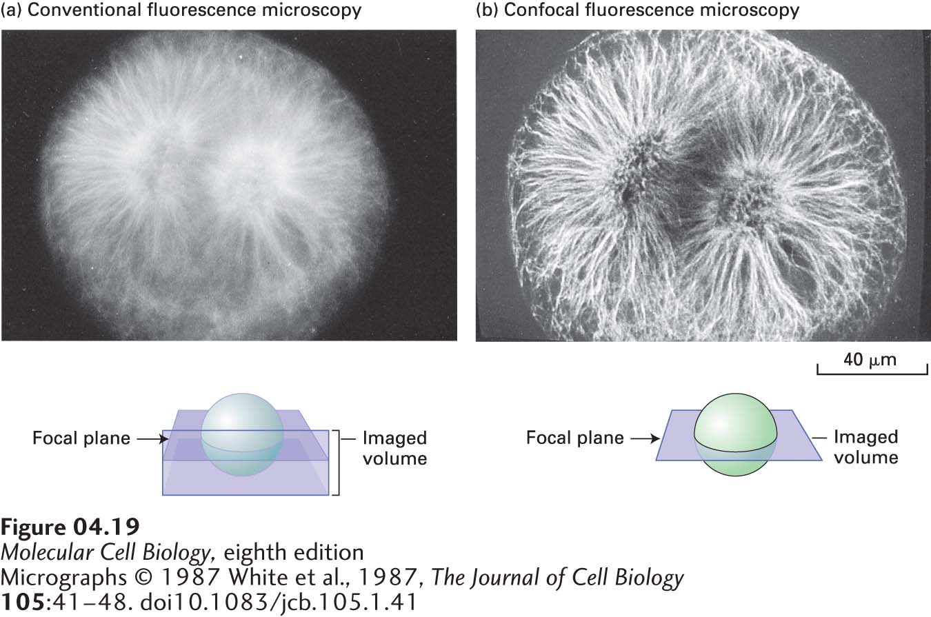
FIGURE 4- 19 Confocal microscopy produces an in- focus optical section through thick cells. A mitotic fertilized egg from a sea urchin (Psammechinus) was lysed with a detergent, exposed to an tubulin antibody, and then exposed to a fluorescein- tagged antibody that binds to the anti- tubulin antibody. (a) When viewed by conventional fluorescence microscopy, the mitotic spindle is blurred. This blurring occurs because background fluorescence is detected from tubulin above and below the focal plane as depicted in the sketch. (b) The confocal microscopic image is sharp, particularly in the center of the mitotic spindle. In this case, fluorescence is detected only from molecules in the focal plane, generating a very thin optical section.
[Micrographs © 1987 White et al., 1987, The Journal of Cell Biology 105:41– 48. doi10.1083/jcb.105.1.41]
[Leave] [Close]