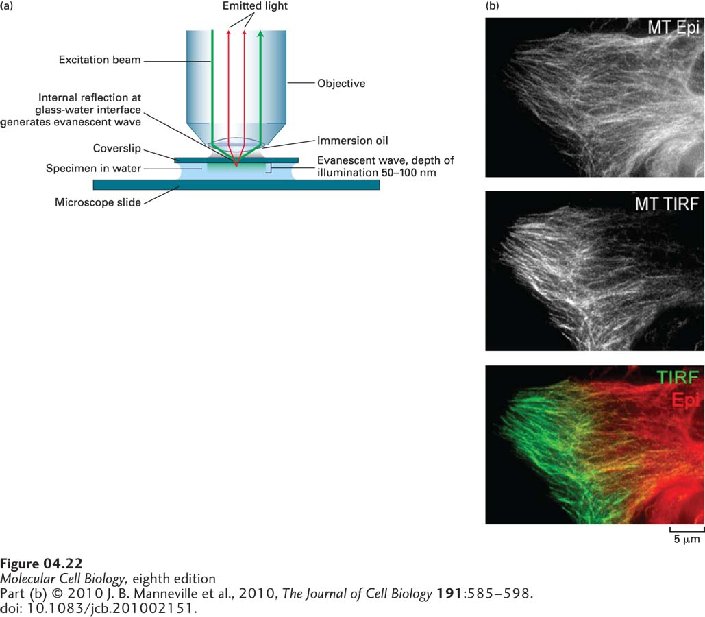
EXPERIMENTAL FIGURE 4- 22 Fluorescent samples in a restricted focal plane can be imaged by total internal reflection fluorescence (TIRF) microscopy. (a) In TIRF microscopy, only about 50– 100 nm of the specimen adjacent to the coverslip is illuminated, so that fluorescent molecules in the rest of the sample are not excited. This limited illumination is achieved by directing the illuminating light at an angle at which it is reflected from the glass- water interface of the coverslip rather than passing through it. Whereas most of the light is reflected, it also generates a very small region of illumination called the evanescent wave (depicted in light green). (b) Immunofluorescence microscopy with tubulin antibody was used to visualize microtubules viewed by conventional fluorescence microscopy (top) and by TIRF (middle), and a merged image was created from the two views (bottom). The two images were collected and false- colored red and green so that the merge could highlight those microtubules that are close to the coverslip (green).
[Part (b) © 2010 J. B. Manneville et al., 2010, The Journal of Cell Biology 191:585– 598. doi: 10.1083/jcb.201002151.]
[Leave] [Close]