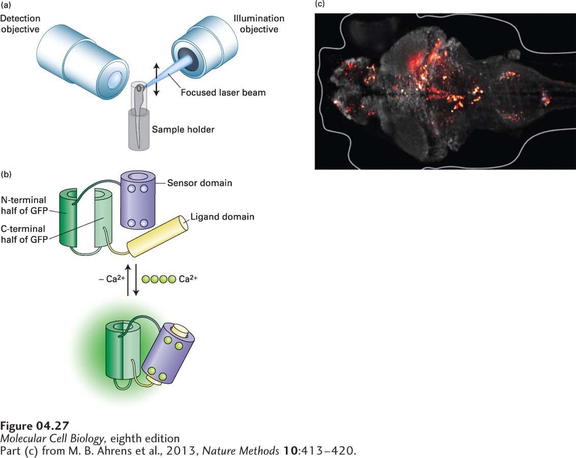
FIGURE 4- 27 Light- sheet microscopy can image rapid events in living tissue. (a) In light- sheet microscopy, a tissue sample is illuminated from the side by a focused laser beam that scans the sample to generate a sheet of light. The sample is observed in the orthogonal direction though the detection objective. To get a three- dimensional image, the illuminating and detection objectives are moved coordinately, taking images throughout the depth of the sample. (b) The Ca2+ biosensor known as GCaMP. This biosensor is made using recombinant DNA techniques to generate a polypeptide to which the N- and C- termini of GFP are fused, and the middle interrupted. On one side of the interruption is the sensor domain, consisting of the Ca2+-binding protein calmodulin. On the other side is the ligand domain, consisting of a target sequence to which calmodulin will bind in the presence of Ca2+. In the absence of Ca2+, the GFP is not functional due to the interruption. In the presence of Ca2+, calmodulin binds four Ca2+ ions and undergoes a conformational change that allows it to bind the ligand domain. This conformational change brings the two parts of GFP into close proximity so that the fluorescent protein is functional. (c) Ca2+ transients, false- colored red, in cells of the brain of a living zebrafish.
[Part (c) from M. B. Ahrens et al., 2013, Nature Methods 10:413– 420.]
[Leave] [Close]