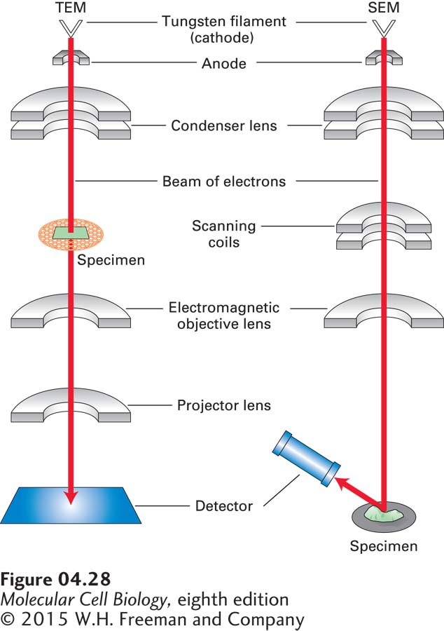
FIGURE 4- 28 In electron microscopy, images are formed from electrons that pass through a specimen or are scattered from a metal- coated specimen. In a transmission electron microscope (TEM, left), electrons are extracted from a heated filament, accelerated by an electric field, and focused on the specimen by a magnetic condenser lens. Electrons that pass through the specimen are focused by a series of magnetic objective and projector lenses to form a magnified image of the specimen on a detector, which may be a fluorescent viewing screen, a photographic film, or a charged- couple- device (CCD) camera. In a scanning electron microscope (SEM), electrons are focused by condenser and objective lenses on a metal- coated specimen. Scanning coils move the beam across the specimen, and electrons scattered from the metal are collected by a photomultiplier tube detector. In both types of microscopes, because electrons are easily scattered by air molecules, the entire column is maintained at a very high vacuum.
[Leave] [Close]