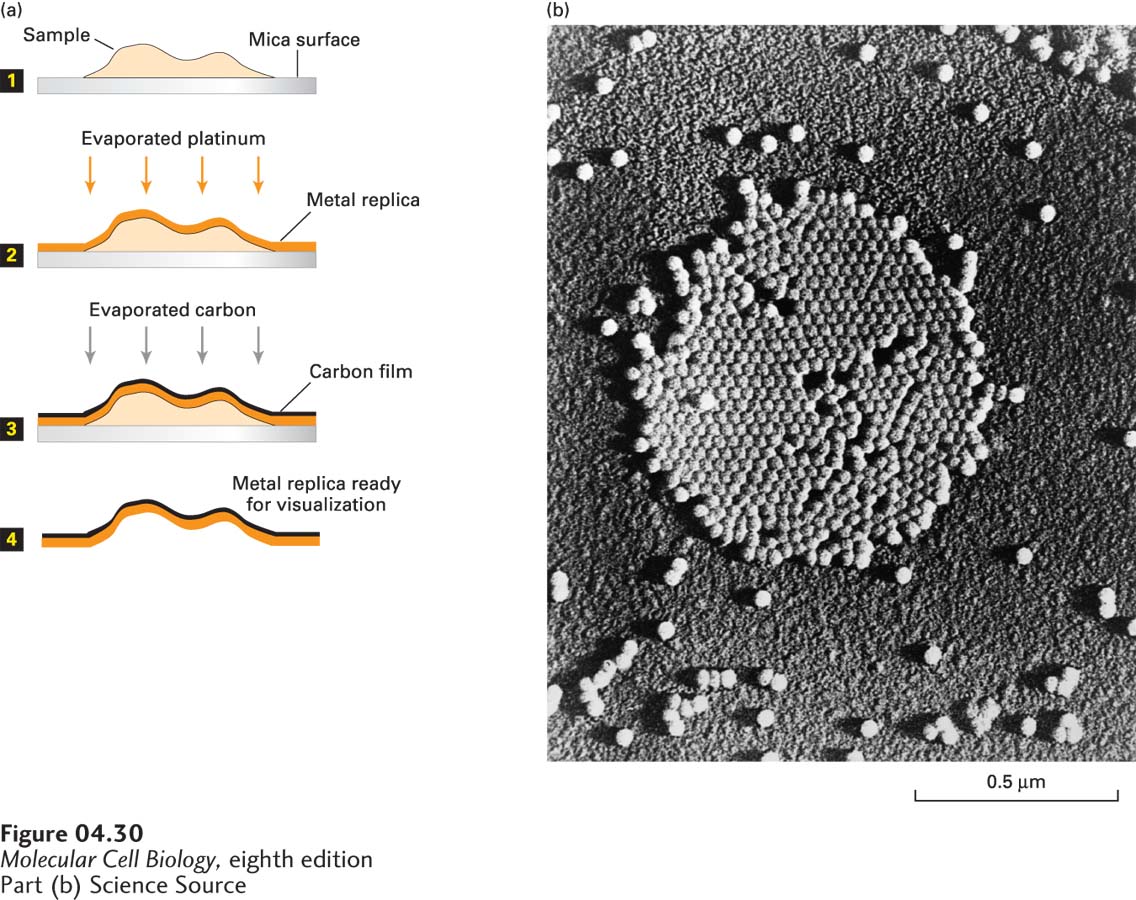
FIGURE 4- 30 Metal shadowing makes surface details on very small objects visible by transmission electron microscopy. (a) The sample is spread on a mica surface and then dried in a vacuum evaporator (step 1). The sample grid is coated with a thin film of a heavy metal, such as platinum or gold, evaporated from an electrically heated metal filament (step 2). To stabilize the replica, the specimen is then coated with a carbon film evaporated from an overhead electrode (step 3). The biological material is then dissolved by acid and bleach (step 4), and the remaining metal replica is viewed in a TEM. In electron micrographs of such preparations, the carbon- coated areas appear light— the reverse of micrographs of simple metal- stained preparations, in which the areas of heaviest metal staining appear the darkest. (b) A platinum- shadowed replica of poliovirus particles.
[Part (b) Science Source]
[Leave] [Close]