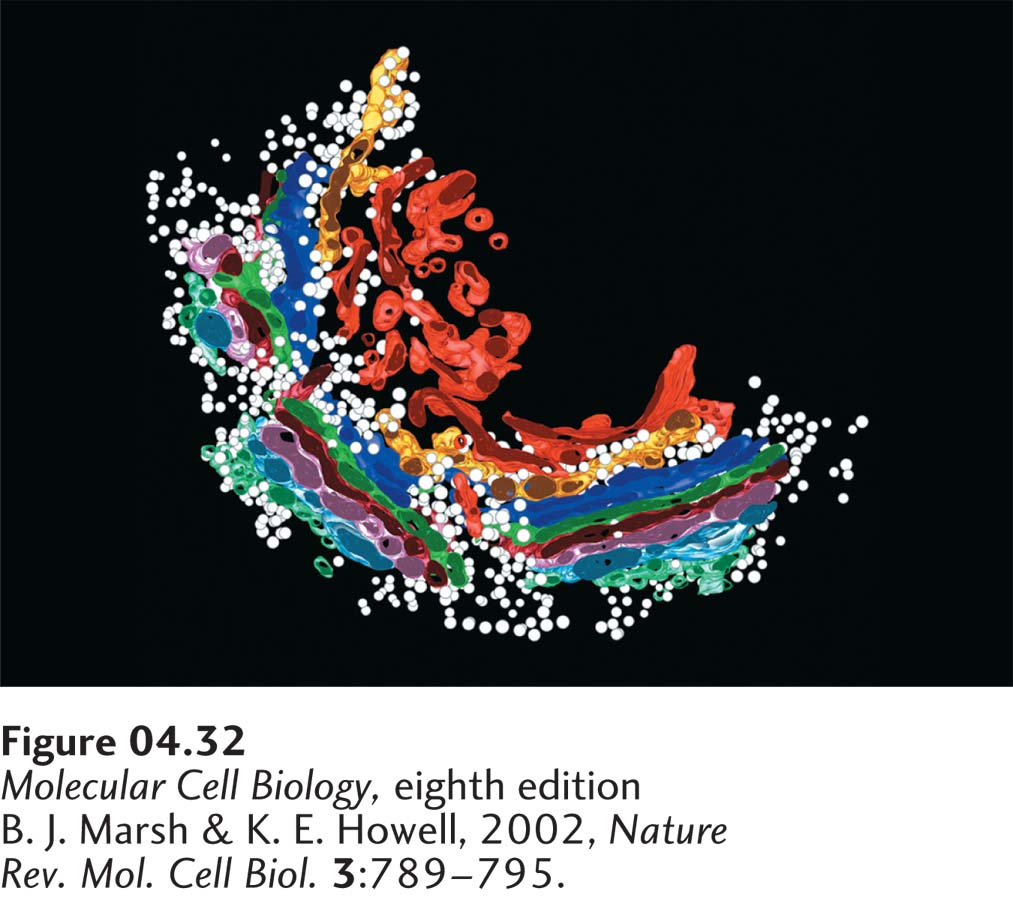
FIGURE 4- 32 Model of the Golgi complex based on three- dimensional reconstruction of electron microscopy images. Transport vesicles (white spheres) that have budded off the rough ER fuse with the cis membranes (light blue) of the Golgi complex. By mechanisms described in Chapter 14, proteins move from the cis region to the medial region and finally to the trans region of the Golgi complex. Eventually, vesicles bud off the trans-Golgi membranes (orange and red); some move to the cell surface and others move to lysosomes. The Golgi complex, like the rough endoplasmic reticulum, is especially prominent in secretory cells.
[B. J. Marsh & K. E. Howell, 2002, Nature Rev. Mol. Cell Biol. 3:789– 795.]
[Leave] [Close]