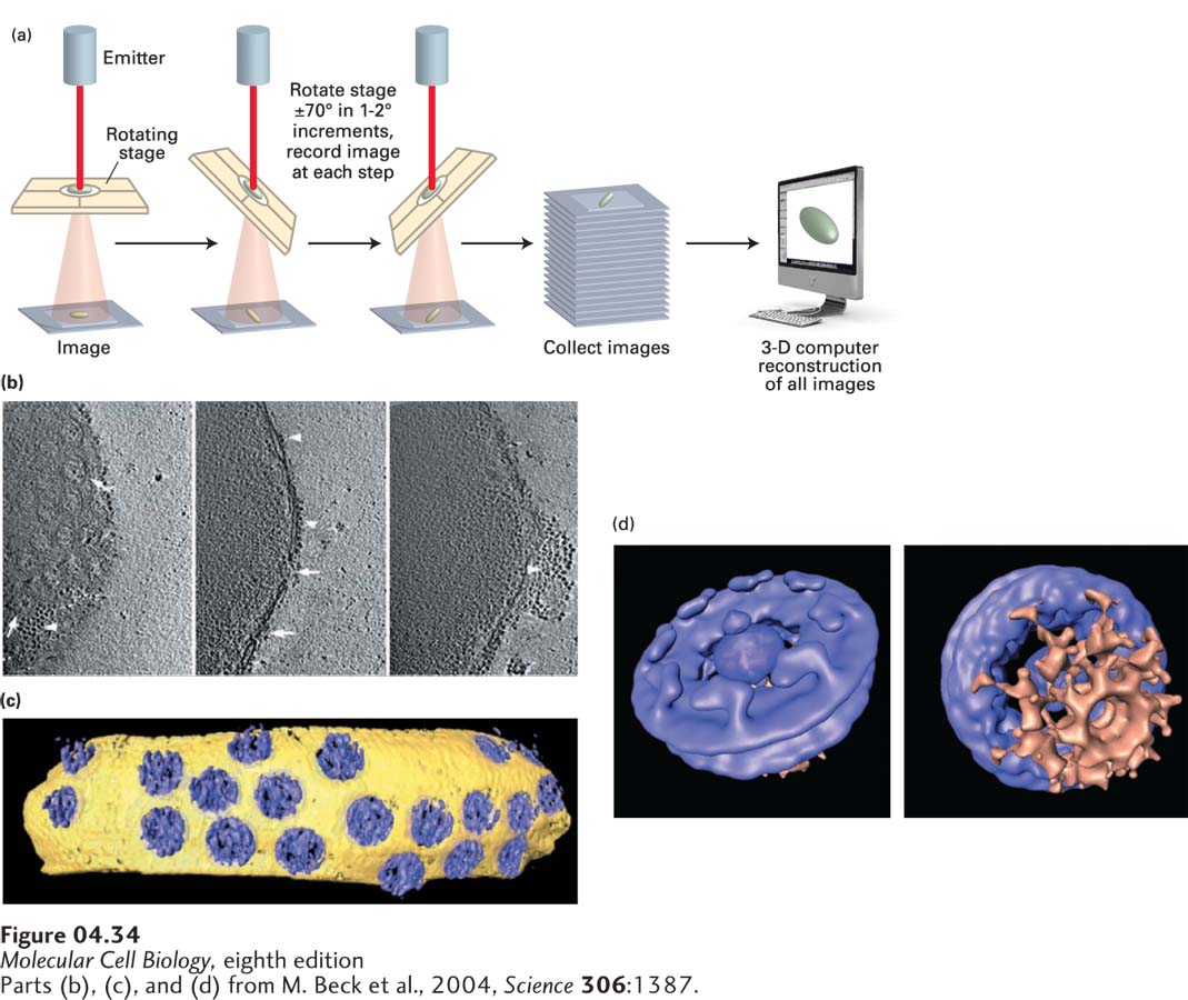
FIGURE 4- 34 Structure of the nuclear pore complex (NPC) imaged by cryoelectron tomography. (a) In cryoelectron tomography, a semicircular series of two- dimensional projection images is recorded from the three- dimensional specimen that is located at the center; the specimen is tilted while the electron optics and detector remain stationary. The three- dimensional structure is then computed from the collected two- dimensional images. (b) Isolated nuclei from the cellular slime mold Dictyostelium discoideum were quick- frozen in liquid nitrogen and maintained in this state as the sample was observed in the electron microscope. The panel shows three sequential tilted images. Different orientations of NPCs (arrows) are shown in top view (left and center) and side view (right). Ribosomes connected to the outer nuclear membrane are visible, as is a patch of rough ER (arrowheads). (c) Computer- generated surface- rendered representation of a segment of the nuclear envelope membrane (yellow) studded with NPCs (blue). (d) By averaging the images of multiple nuclear pores, much more detail can be discerned. See S. Nickell et al., 2006, Nature Rev. Mol. Cell Biol. 7:225.
[Parts (b), (c), and (d) from M. Beck et al., 2004, Science 306:1387.]
[Leave] [Close]