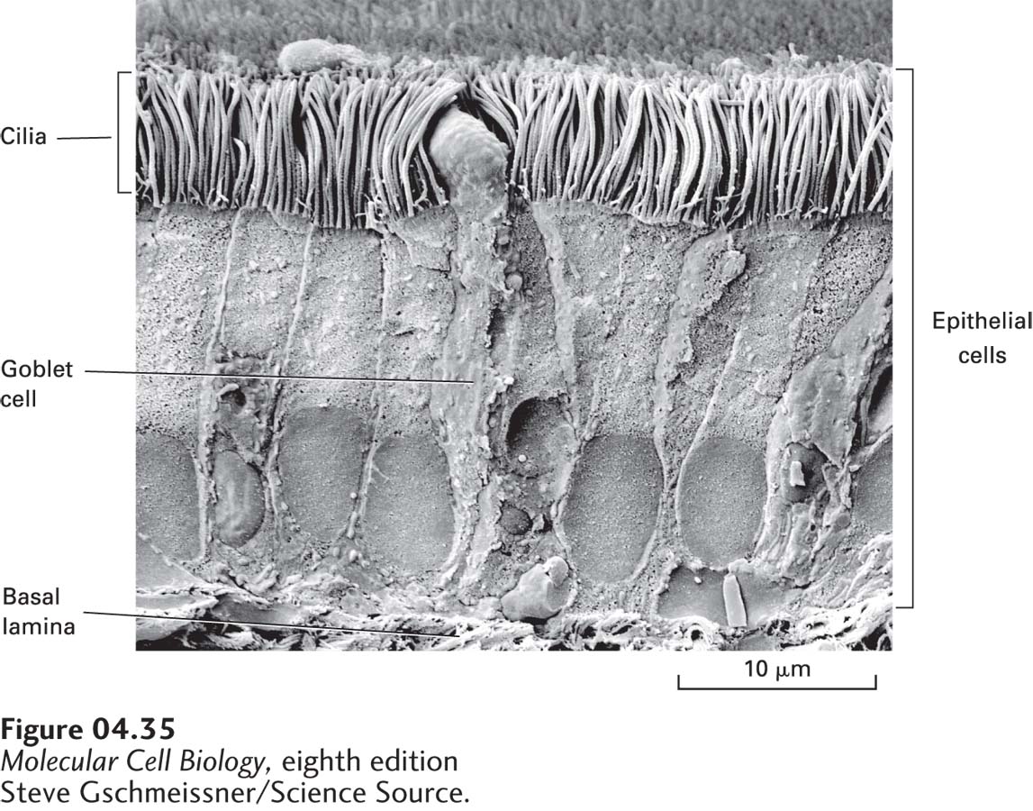
FIGURE 4- 35 Scanning electron microscopy (SEM) produces a three- dimensional image of the surface of an unsectioned specimen. Seen here is an SEM image of cells of the trachea. In the middle is a goblet cell, which secretes mucus. On either side of the goblet cell are epithelial cells with abundant cilia on their apical surfaces.
[Steve Gschmeissner/Science Source.]
[Leave] [Close]