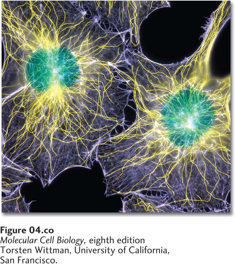
Fluorescence microscopy showing the locations of DNA (green), microtubules (yellow), and microfilaments (purple) in two cultured cells. The cells were chemically fixed and then rendered permeable to antibodies using a gentle detergent. Microtubules were stained with an antibody to tubulin; microfilaments were stained with a labeled toxin, phalloidin, that binds selectively to F- actin; and DNA was visualized with a DNA- binding dye.
[Torsten Wittman, University of California, San Francisco.]
[Leave] [Close]