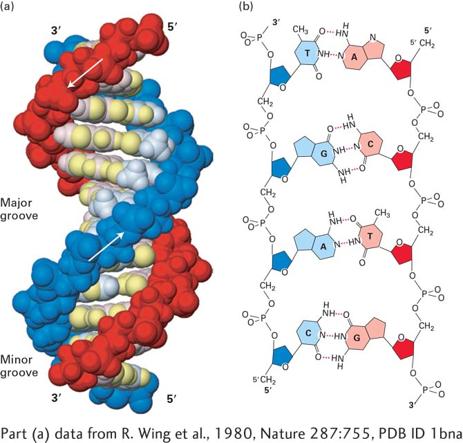
FIGURE 5- 3 The DNA double helix. (a) Space- filling model of B DNA, the most common form of DNA in cells. The bases (light shades) project inward from the sugar- phosphate backbones (dark red and blue) of each strand, but their edges are accessible through major and minor grooves. Arrows indicate the 5′→3′ direction of each strand. Hydrogen bonds between the bases are in the center of the structure. The major and minor grooves are lined by potential hydrogen bond donors and acceptors (highlighted in yellow). (b) Chemical structure of DNA double helix. This extended schematic shows the two sugar- phosphate backbones and hydrogen bonding between the Watson- Crick base pairs, A·T and G·C. See R. E. Dickerson, 1983, Sci. Am. 249:94.
[Part (a) data from R. Wing et al., 1980, Nature 287:755, PDB ID 1bna.]
[Leave] [Close]