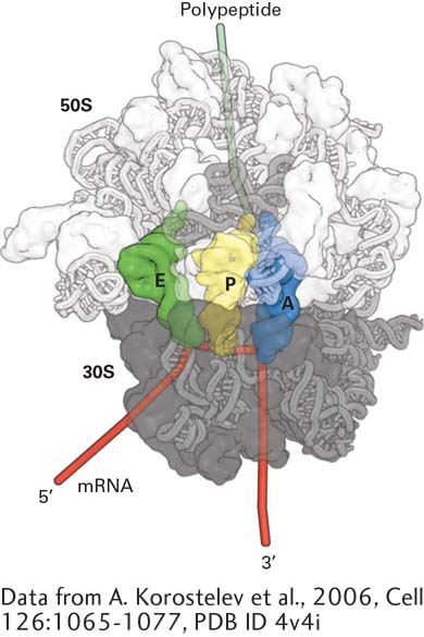
FIGURe 5- 22 Structure of the bacterial ribosome. Model of the T. thermophilus ribosome viewed along the interface between the large (50S) and small (30S) subunits. The 16S rRNA and proteins in the small subunit are dark gray. RNA is depicted as a tube model and protein surfaces are shown; the 23S rRNA and proteins in the large subunit are light gray; and the 5S rRNA is an intermediate shade of gray. The surface of the ribosome is made partially transparent to display the positions of tRNAs in the A, P, and E sites. Note that the ribosomal proteins are located primarily on the surface of the ribosome.
[Data from A. Korostelev et al., 2006, Cell 126:1065- 1077, PDB ID 4v4i.]
[Leave] [Close]